. 2009 Apr;174(4):1435-42.
doi: 10.2353/ajpath.2009.080560. Epub 2009 Feb 26.

Large CTG repeats trigger p16-dependent premature senescence in myotonic dystrophy type 1 muscle precursor cells

Affiliations

Large CTG repeats trigger p16-dependent premature senescence in myotonic dystrophy type 1 muscle precursor cells

Anne Bigot et al. Am J Pathol. 2009 Apr.

Abstract

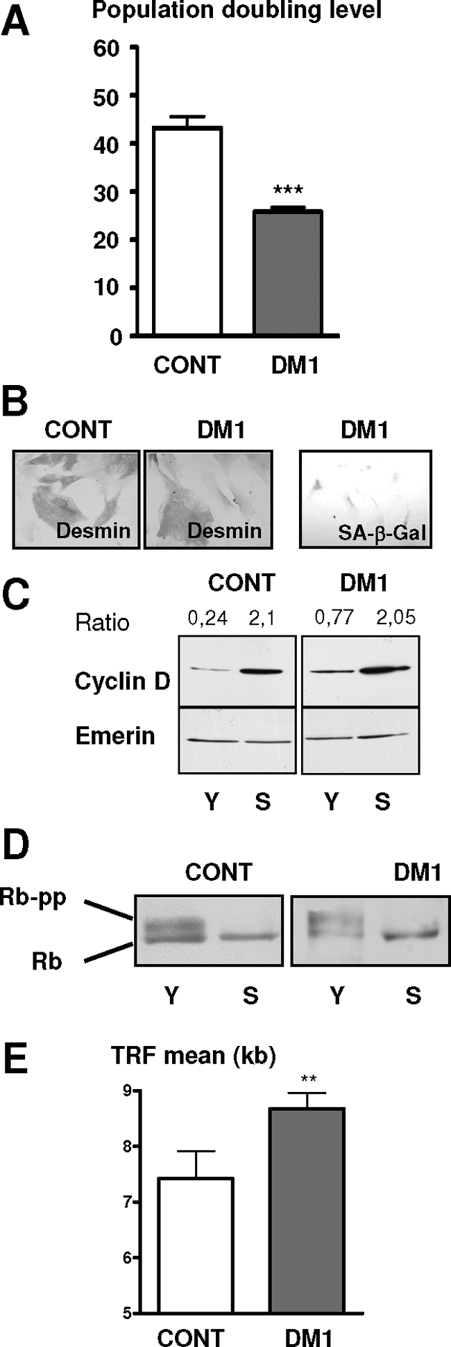
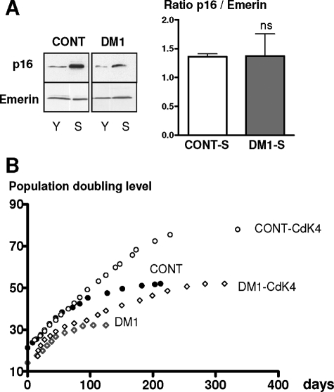
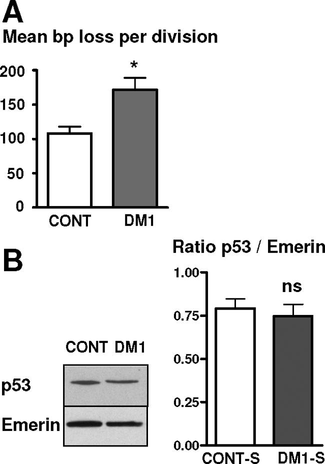
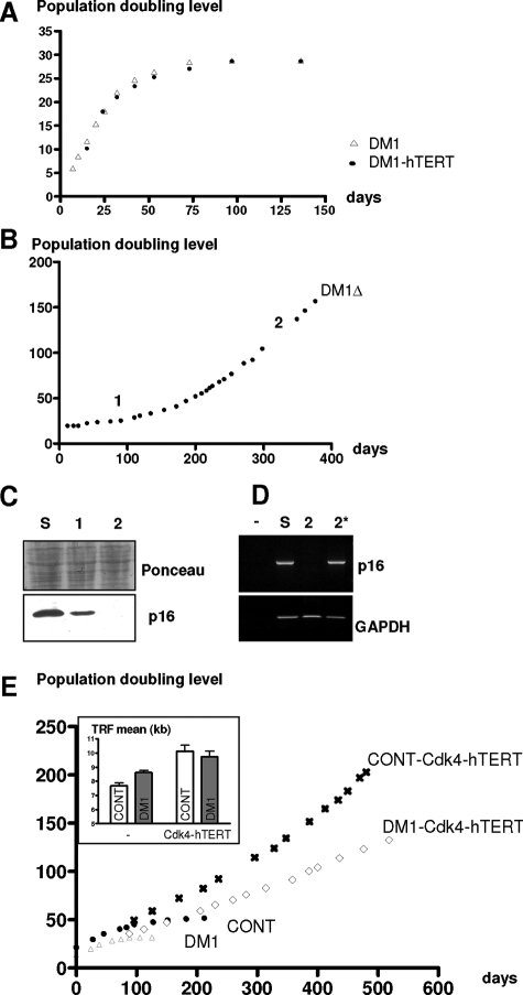
A CTG repeat amplification is responsible for the dominantly inherited neuromuscular disorder, myotonic dystrophy type 1 (DM1), which is characterized by progressive muscle wasting and weakness. The expanded (CTG)n tract not only alters the myogenic differentiation of the DM1 muscle precursor cells but also reduces their proliferative capacity. In this report, we show that these muscle precursor cells containing large CTG expansion sequences have not exhausted their proliferative capacity, but have entered into premature senescence. We demonstrate that an abnormal accumulation of p16 is responsible for this defect because the abolition of p16 activity overcomes early growth arrest and restores an extended proliferative capacity. Our results suggest that the accelerated telomere shortening measured in DM1 cells does not contribute to the aberrant induction of p16. We propose that a cellular stress related to the amplified CTG repeat promotes premature senescence mediated by a p16-dependent pathway in DM1 muscle precursor cells. This mechanism is responsible for the reduced proliferative capacity of the DM1 muscle precursor cells and could participate in both the impaired regeneration and atrophy observed in the DM1 muscles containing large CTG expansions.


Figures

Figure 1
Figure 1
DM1 satellite cells stop dividing by a premature senescence pathway. A: Mean of the population doubling level of DM1 and control satellite cells isolated from muscles of three DM1 patients with large CTG repeats (>2000) and three age-matched and nonaffected individuals, respectively. B: Immunocytochemistry using anti-desmin antibody and revealed by peroxidase was performed on senescent control and premature growth-arrested DM1 cells. Flattened and enlarged morphologies are present in both arrested DM1 cells and control senescent cells. Senescent-associated ß-galactosidase activity (SA-ß-Gal) was observed on arrested DM1 cells. C: Western blot analyses of cyclin D in the early (Y) and late (S) stages of their proliferative lifespan. The results were normalized to the expression of the nuclear protein Emerin. Levels of cyclin D were increased in both DM1- and control-arrested cells. D: Western blot analyses of Rb in the early (Y) and late (S) stages of their proliferative lifespan. The hyperphosphorylated form of Rb is absent in both controls and DM1-arrested cells. E: Mean length of telomeric restriction fragments (TRFs) measured on DM1 and control satellite cells at proliferative arrest. DM1 satellite cells stop dividing with longer telomeres than controls. (**P < 0.01, ***P < 0.001).
Figure 2
Figure 2
Premature growth arrest of DM1 satellite cells is dependent on the p16 pathway. A: p16 levels in control and DM1 satellite cells at early (Y) and late (S) stages in their proliferative lifespan were determined by Western blot. Means of the p16/Emerin ratio in control and DM1 senescent cells were presented on the histogram. Control- and DM1-arrested cells present the same level of expression of p16, ns: not significant. B: Lifespan plots of control and DM1 populations transduced with or without Cdk4. Transduction of Cdk4 gene allows to bypass the arrest observed in nontransduced cells.
Figure 3
Figure 3
Deregulation of the telomere homeostasis in DM1 satellite cells. A: Mean length of telomeric DNA in bp lost per division in cultures of control and DM1 satellite cells. DM1 cells lost more bp per division than control cells. B: Western blot analyses of the level of p53 in control and DM1 satellite cells at the end of their lifespan. Means of the p53/Emerin ratio in control- and DM1-arrested cells are presented on the histogram. *P < 0.05, ns: not significant.
Figure 4
Figure 4
Immortalization of DM1 satellite cells. A: Lifespan plots of DM1 satellite cells transduced with or without hTERT. hTERT-transduced cells were not able to bypass the premature arrest observed in DM1 satellite cells. B: Lifespan plot of a clone of cells (DM1Δ) that emerged from a DM1 satellite cell population transduced with hTERT. DM1Δ cells were immortalized. C: Western blot analyses of p16 in senescent DM1 cells (lane S), in the senescent population with emerging DM1Δ cells (lane 1), and in the DM1Δ after several divisions (lane 2). The two points (1 and 2) are also indicated on the DM1Δ lifespan curve shown above. D: Expression of p16 and GAPDH mRNA analyzed by RT-PCR: negative control (lane −), senescent DM1 satellite cells (lane S), DM1Δ (lane 2), DM1Δ treated with 5-aza-2′deoxycytidine (lane 2*). E: Lifespan plots of DM1 satellite cells transduced with or without Cdk4 and hTERT. Cdk4- and hTERT-transduced DM1 cells were immortalized. Effect of hTERT on the telomere length is presented in the inset. hTERT stabilized the telomere length of the cells ∼10 kb.

References

    1. Harper PS. London: W.B. Saunders,; Myotonic Dystrophy. (ed 3) 2004:pp. 17–45.
    1. Aslanidis C, Jansen G, Amemiya C, Shutler G, Mahadevan M, Tsilfidis C, Chen C, Alleman J, Wormskamp NG, Vooijs M, Buxton J, Johnson K, Smeets JM, Lennon G, Carrano AV, Korneluk R, Wieringa B, de Jong P. Cloning of the essential myotonic dystrophy region and mapping of the putative defect. Nature. 1992;355:548–551. - PubMed
    1. Brook JD, McCurrach ME, Harley HG, Buckler AJ, Church D, Aburatani H, Hunter K, Stanton VP, Thirion JP, Hudson T, Sohn R, Zemelman B, Snell RG, Rundle SA, Crow S, Davies J, Shelbourne P, Buxton J, Jones C, Juvonen V, Johnson K, Harper P, Shaw DJ, Housman D. Molecular basis of myotonic dystrophy: expansion of a trinucleotide (CTG) repeat at the 3′ end of a transcript encoding a protein kinase family member. Cell. 1992;68:799–808. - PubMed
    1. Fu YH, Pizzuti A, Fenwick RG, Jr, King J, Rajnarayan S, Dunne PW, Dubel J, Nasser GA, Ashizawa T, de Jong P, Wieringa B, Korneluk R, Perryman MB, Epstein HF, Caskey CT. An unstable triplet repeat in a gene related to myotonic muscular dystrophy. Science. 1992;255:1256–1258. - PubMed
    1. Mahadevan M, Tsilfidis C, Sabourin L, Shutler G, Amemiya C, Jansen G, Neville C, Narang M, Barcelo J, O'Hoy K, Leblond S, Earl-Macdonald J, de Jong P, Wieringa B, Korneluk R. Myotonic dystrophy mutation: an unstable CTG repeat in the 3′ untranslated region of the gene. Science. 1992;255:1253–1255. - PubMed

Publication types

MeSH terms

Substances