. 2009 Mar;155(Pt 3):979-988.
doi: 10.1099/mic.0.024539-0.

Evaluation of procedures for outer membrane isolation from Campylobacter jejuni

Affiliations

Evaluation of procedures for outer membrane isolation from Campylobacter jejuni

Rhonda I Hobb et al. Microbiology (Reading). 2009 Mar.

Abstract

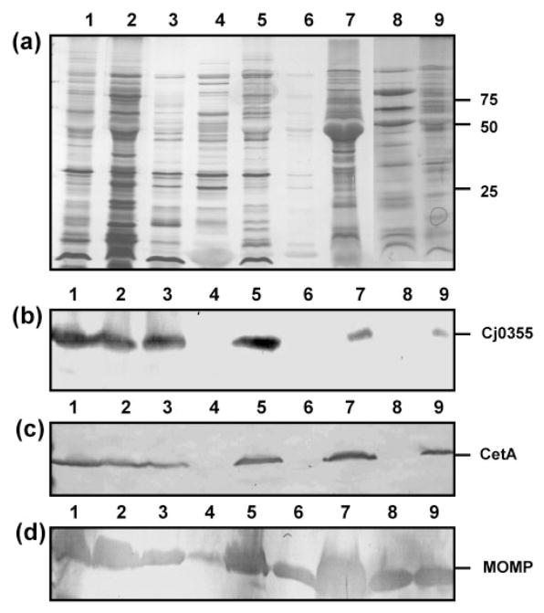
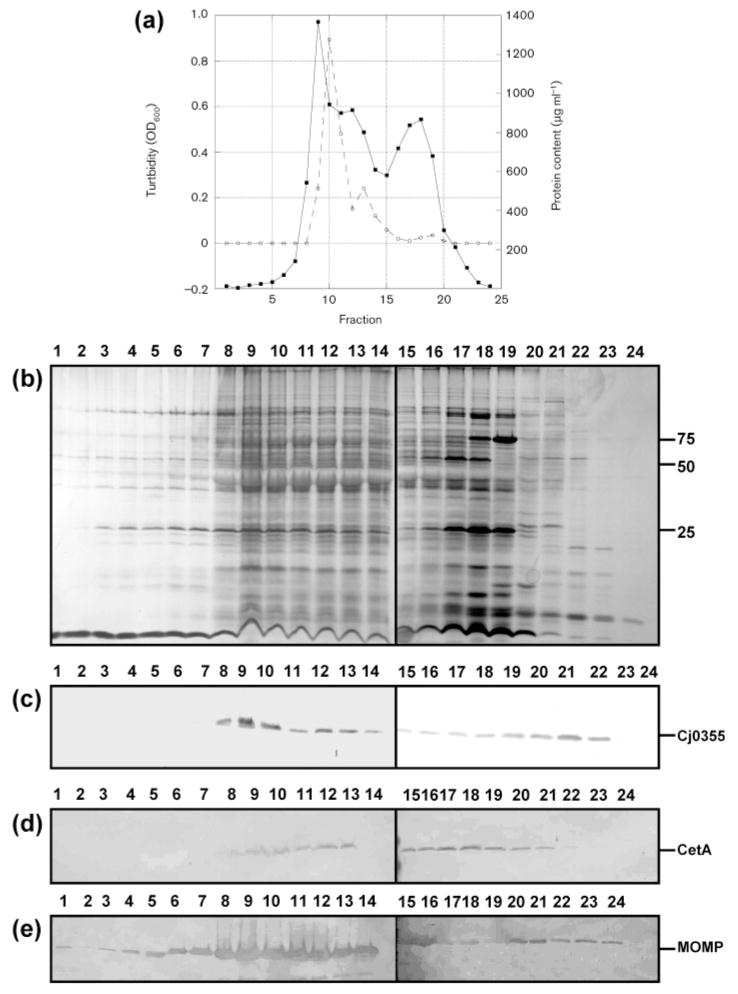
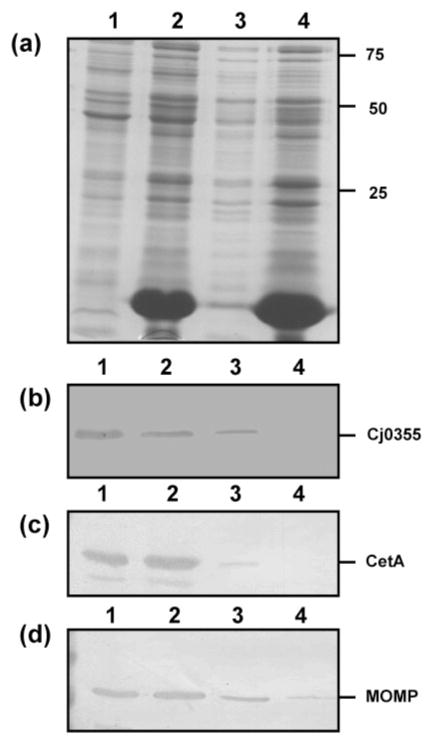
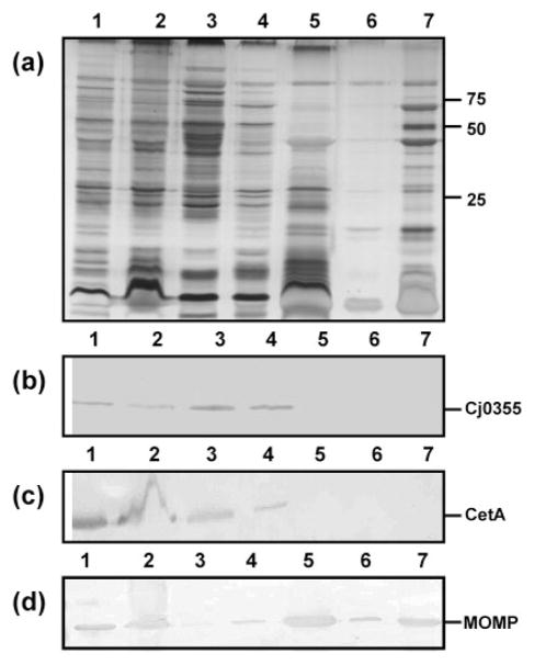
Although infection with Campylobacter jejuni is one of the leading causes of gastroenteritis worldwide, relatively little is known about the factors that are required to elicit a protective immune response. The need for a vaccine against this pathogen is well recognized and a number of vaccine candidates have been tested with varying degrees of success; however, there is still a lack of a suitable vaccine. To gain a better understanding of the outer-membrane protein components of this organism, a 'gold standard' method to purify the outer membrane is needed. Therefore, we attempted to develop a robust and reliable method which resulted in a pure outer-membrane fraction. A total of nine methodologies were examined and analysed by SDS-PAGE and immunoblotting using subcellular markers for the cytoplasm, cytoplasmic membrane and outer membrane. We found that glycine extraction, differential detergent extraction using Triton X-100, serial extraction using 1 M Tris pH 7, spheroplasting by lysozyme and sonication, and carbonate extraction did not produce pure outer-membrane preparations. However, we identified three methods that provided outer-membrane fractions free from subcellular contamination. Isopycnic centrifugation using a 30-60 % sucrose gradient produced seven fractions free from cytoplasmic or cytoplasmic membrane contamination; however, these fractions did not correspond as well as expected with the typical outer-membrane-associated peak (e.g. Escherichia coli or Salmonella). The spheroplast method using lysozyme alone also resulted in pure outer-membrane fraction, as did carbonate washing of this sample. The extraction of outer membranes using N-lauroylsarcosine (Sarkosyl) produced the purest and most reproducible sample. These outer-membrane preparations will be useful for future studies aimed at identifying C. jejuni surface proteins as vaccine components.


Figures

Fig. 1
Fig. 1
Outer-membrane fractions of C. jejuni strain 81–176 from nine purification methods. (a) Silver-stained SDS-PAGE analysis of OMP preparations (5 μg). Lanes: 1, glycine extraction; 2, differential detergent extraction using Triton X-100; 3; serial extraction using 0.1 M Tris/HCl, pH 7.8; 4, spheroplasts isolated using lysozyme treatment; 5, spheroplasts isolated using sonication and lysozyme treatment; 6, carbonate washed spheroplasts; 7, carbonate extraction; 8, Sarkosyl extraction; 9, sucrose gradient fraction 14. Molecular masses are indicated on the right (in kDa). (b–d) Immunoblots of the outer-membrane preparations using the cytoplasmic marker anti-Cj0355 (b), the inner-membrane marker anti-CetA (c) and the outer-membrane marker anti-MOMP (d). All antibodies were used at 1: 1000 dilution. Loading for panels (b)–(d) is the same as for panel (a).
Fig. 2
Fig. 2
Analysis of fractions obtained from the sucrose gradient of C. jejuni strain 81–176. (a) Graphical representation of the turbidity measured as OD600 (solid line) and protein content measured by BCA assay (dashed line) for fractions 1–24. (b) Silver-stained SDS-PAGE gel of sucrose gradient fractions. Lanes 1–24, fractions 1–24 (10 μl). Molecular masses are indicated on the right (in kDa). (c–e) Immunoblots of sucrose gradient fractions 1–24 using the cytoplasmic marker anti-Cj0355 (c), the inner-membrane marker anti-CetA (d) and the outer-membrane marker anti-MOMP (e). All antibodies were used at 1: 1000 dilution. Loading for panels (c)–(e) is the same as for panel (b).
Fig. 3
Fig. 3
Analysis of the preparative steps for spheroplasts of C. jejuni strain 81–176 using the lysozyme method. (a) Silver-stained SDS-PAGE gel of the preparative steps. Lanes: 1, whole-cell lysate; 2, post-lysozyme treatment supernatant; 3, inner membrane isolated from spheroplasts; 4, supernatant following centrifugation to remove unlysed cells (contains mostly outer membrane). Ten microlitres of each of the preparative steps was loaded per lane. Molecular masses are indicated on the right (in kDa). (b–d) Immunoblots of the lysozyme spheroplast preparatory samples using the cytoplasmic marker anti-Cj0355 (b), the inner-membrane marker anti-CetA (c) and the outer-membrane marker anti-MOMP (d). All antibodies were used at 1: 1000 dilution. Loading for panels (b)–(d) is the same as for panel (a).
Fig. 4
Fig. 4
Analysis of the preparative steps for the OMPs of C. jejuni strain 81–176 using Sarkosyl. (a) Silver-stained SDS-PAGE gel of the preparative steps. Lanes: 1, whole-cell lysate (10 μl); 2, lysate following French pressure lysis of whole cells (10 μl); 3, ultracentrifuge supernatant from first wash with 10 mM HEPES, pH 7.4 (10 μl); 4, ultracentrifuge supernatant from second wash with 10 mM HEPES, pH 7.4 (10 μl); 5, supernatant following Sarkosyl incubation (10 μl); 6, supernatant following final wash with 10 mM HEPES, pH 7.4 (10 μl); 7, Sarkosyl-insoluble proteins (OMPs) (5 μg). Molecular masses are indicated on the right (in kDa). (b–d) Immunoblots using the cytoplasmic marker anti-Cj0355 (b), the inner-membrane marker anti-CetA (c) and the outer-membrane marker anti-MOMP (d). All antibodies were used at 1: 1000 dilution.

Similar articles

Cited by

References

    1. Alahari A, Saint N, Campagna S, Molle V, Molle G, Kremer L. The N-terminal domain of OmpATb is required for membrane translocation and pore-forming activity in mycobacteria. J Bacteriol. 2007;189:6351–6358. - PMC - PubMed
    1. Albert MJ, Haridas S, Steer D, Dhaunsi GS, Smith AI, Adler B. Identification of a Campylobacter jejuni protein that cross-reacts with cholera toxin. Infect Immun. 2007;75:3070–3073. - PMC - PubMed
    1. Allos BM. Campylobacter infections: update on emerging issues and trends. Clin Infect Dis. 2001;32:1201–1206. - PubMed
    1. Baqar S, Applebee LA, Bourgeois AL. Immunogenicity and protective efficacy of a prototype Campylobacter killed whole-cell vaccine in mice. Infect Immun. 1995;63:3731–3735. - PMC - PubMed
    1. Bayer ME. Areas of adhesion between wall and membrane of Escherichia coli. J Gen Microbiol. 1968;53:395–404. - PubMed

Publication types

MeSH terms

LinkOut - more resources