. 2009 Feb;5(2):e1000309.
doi: 10.1371/journal.ppat.1000309. Epub 2009 Feb 27.

Export of a Toxoplasma gondii rhoptry neck protein complex at the host cell membrane to form the moving junction during invasion

Affiliations

Export of a Toxoplasma gondii rhoptry neck protein complex at the host cell membrane to form the moving junction during invasion

Sébastien Besteiro et al. PLoS Pathog. 2009 Feb.

Abstract

One of the most conserved features of the invasion process in Apicomplexa parasites is the formation of a moving junction (MJ) between the apex of the parasite and the host cell membrane that moves along the parasite and serves as support to propel it inside the host cell. The MJ was, up to a recent period, completely unknown at the molecular level. Recently, proteins originated from two distinct post-Golgi specialised secretory organelles, the micronemes (for AMA1) and the neck of the rhoptries (for RON2/RON4/RON5 proteins), have been shown to form a complex. AMA1 and RON4 in particular, have been localised to the MJ during invasion. Using biochemical approaches, we have identified RON8 as an additional member of the complex. We also demonstrated that all RON proteins are present at the MJ during invasion. Using metabolic labelling and immunoprecipitation, we showed that RON2 and AMA1 were able to interact in the absence of the other members. We also discovered that all MJ proteins are subjected to proteolytic maturation during trafficking to their respective organelles and that they could associate as non-mature forms in vitro. Finally, whereas AMA1 has previously been shown to be inserted into the parasite membrane upon secretion, we demonstrated, using differential permeabilization and loading of RON-specific antibodies into the host cell, that the RON complex is targeted to the host cell membrane, where RON4/5/8 remain associated with the cytoplasmic face. Globally, these results point toward a model of MJ organization where the parasite would be secreting and inserting interacting components on either side of the MJ, both at the host and at its own plasma membranes.


Conflict of interest statement

The authors have declared that no competing interests exist.

Figures

Figure 1
Figure 1. Identification of an additional moving junction complex member, RON8.
(A) Isolation of RON4-associated proteins by immuno-affinity column. Eluate from the purification of a tachyzoite lysate on T5 4H1 immunosorbent was resolved by 8% SDS-PAGE and stained with colloidal blue. Bands taken for mass spectrometry analysis are numbered on the left, and peptides identified are listed in Table S1. Bands previously identified as RON2, RON4, and RON5 are indicated, as well as band corresponding to immunoglobulin (Ig). Molecular mass standards are indicated on the right. (B) Co-localisation by IFA of RON8 (Tw2001) with RON4 to the rhoptry neck within intracellular tachyzoites fixed and permeabilized with methanol. The absence of reactivity of the pre-immune serum is presented in the lower panel as a negative control. (C) Lysate: Western blot with anti- Tw2001 of a Toxoplasma lysate run in non-reduced condition. (pi: pre-immune serum). IP αTw2001: Co-IP of RON8, RON4, and AMA1 using anti-RON8 antibody. Proteins isolated on an anti-RON8 immuno-affinity column were separated on SDS-PAGE in reduced condition, transferred to nitrocellulose and probed with anti-mouse secondary antibody conjugate alone (Conj), with T5 4H1 mAb (anti-R4) or with CL22 mAb (anti-AMA1). Proteins of interest are shown by arrows; * denotes a non-specific band. Molecular mass standards are indicated on the left. (D) Schematic representation of the MJ proteins with predicted features such as TM domains, as predicted by ConPred II (http://bioinfo.si.hirosaki-u.ac.jp/~ConPred2/), and putative SUB2 cleavage sites (red arrowheads). Regions used as recombinant proteins for the production of the specific antibodies described in Text S1 are underlined.
Figure 2
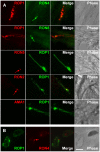
Figure 2. IFA detection of RON2, RON4, RON5, and RON8 in the rhoptry neck, and at the MJ.
Scale bar = 5 µm. (A) Co-localisation of RON2,4,5,8 to the rhoptry neck within intracellular tachyzoites growth overnight on HFF cells, fixed, and permeabilized with methanol. Identical results were obtained with anti-RON4n and anti-RON2n (not shown). Arrow indicates RON4 present in the PV as shown previously . (B) RON8, RON5, and RON4, but not RON2, can be detected at the ring of the MJ during invasion. Phase contrast and IFA of parasites show that mAb anti-RON4, polyclonal anti-RON5, and polyclonal anti-RON8 label the ring corresponding to the MJ in invading parasites permeabilized with 0.05% saponin. The ring was not detected using anti-RON2c (lower panel). (C) As other junctional RONs, RON2 can be detected at the junction in cytD-treated tachyzoites attached to the surface of the host cell. Evacuoles expanding within the host cell cytosol from the site of parasite attachment are labelled with anti-ROP1. RON2 (labelled with anti-RON2c, arrow), RON4, and RON8 are found associated with the tip of the parasite; RON2 is shown to be colocalizing with RON4 in the lower panel.
Figure 3
Figure 3. Interactions between members of the junctional complex.
(A) Western blot analysis in reduced condition of MJ members co-immunoprecipitated in various detergent conditions (1% NP40 or 0.6% SDS) with the antibody mentioned on top and probed with the antibodies mentioned below or probed with secondary antibody conjugate alone (Conj). The first panel corresponds to the Toxoplasma lysate used for the IP and probed with the antibody mentioned below. Molecular mass standards for all the panels are indicated on the left. (B) Co-immunoprecipitation in two detergent conditions of MJ proteins after metabolic labelling, with specific antibodies mentioned below. Metabolic labelling was done with [35S] methionine/cysteine for 15 min on intracellular parasites followed by one hour of culture without radioactivity. Cells were lysed either in 1% NP40 (left panel) or 0.6% SDS (right panel), and proteins were immunopurified using specific antibodies directed against each member of the MJ complex. The IP products were revealed by autoradiography after separation by SDS-PAGE in reduced conditions. MJ proteins positions are shown on the right. Although the profiles in NP40 conditions appear similar, these lanes migrated on independent gels, thus the positions of the RONs are not exactly the same and the molecular mass standards are only indicative.
Figure 4
Figure 4. MJ proteins are processed and can associate as non-mature forms.
(A) Pulse-chase metabolic labelling and IP with specific antibodies showing processing of MJ proteins. T. gondii-infected fibroblasts were labelled for 15 min with [35S] methionine/cysteine and either harvested (P, pulse) or chased for 1 h (C, chase). Then, the 0.6% SDS lysates were immunoprecipitated with the antibody indicated below, and products were run in SDS-PAGE in reduced (R) or non-reduced (NR) conditions before autoradiography. Proteins and their pro-forms are annotated. Molecular mass standards are indicated. (B) Similar analysis in the presence of BFA shows that maturation occurs in a post-Golgi compartment and that non-mature forms are recovered together by IP. Pulse-chase was done as in (A), with or without BFA, and IP were performed on cells lysed in 1% NP40. In the presence of BFA, the same profile was obtained in the pulse and the chase, indicating that pro-RONs can assemble as immature forms. Moreover, proAMA1 could also associate with the RONs complex (lane corresponding to the pulse in the right panel). (C) Biosynthesis of RONs and MICs are asynchronous. Double IFA with anti-proRON8 and anti-proMIC3 antibodies (upper panels) or anti-RON4 and anti-proM2AP antibodies (lower panels) on intracellular parasites fixed with 4% PAF and permeabilized with Triton X-100 label tachyzoites in distinct vacuoles (coloured arrows), at different stages of endodyogeny.
Figure 5
Figure 5. Association of RONs with the host cell plasma membrane.
(A) A RON spot persists on cells after abortive interaction with a parasite in the presence of Cyt-D. Dual IFA were done with anti-RONs and anti-ROP1 on invading parasites stopped by Cyt-D, fixed, and permeabilized with saponin. The anti-ROP1 labeled the evacuoles network resulting from the secretion of rhoptries. The absence of a parasite at the site of secretion of ROP1 and the detection of a single dot with all of the anti-RONs (arrowed), but not with anti-AMA1, indicated that RONs could associate with the host cell membrane. (B) IFA of HFF cells pulse-invaded for 15 min, permeabilized with saponin, and incubated with both the rabbit serum anti-ROP1 and the mAb anti-RON4. The image shows two vacuoles labelled with anti-ROP1, one containing a parasite (right) and the other being empty (left). The arrow indicates the presence of the junctional protein RON4 on the empty vacuole. Scale bar = 5 µm.
Figure 6
Figure 6. RON4, RON5, and RON8 are exposed on the cytosolic face of the host cell plasma membrane.
(A) Differential solubilisation with streptolysin (schematized above) allowed for the specific labelling by IFA of epitopes exposed on the external face of PV membrane. Anti-GRA5Nt and anti-HA9 antibodies served as controls of external and internal PV sides, respectively, in a GRA5-Ct-HA9–tagged transgenic cell line. SAG1 served as a specific control for both extracellular tachyzoites and absence of permeabilization of the PVM in streptolysin-treated cells. RON4, RON5, and RON8 were detected with specific antibody on streptolysin-treated cells, indicating that they are exposed to the cytoplasmic face of the PVM. (B) Glass beads pre-loading of host cells with MJ-specific antibodies and IFA after subsequent invasion by tachyzoites reveals that RON4, RON5, and RON8 are exposed on the cytosolic face of the host cell (arrows). Cells were loaded by antibodies directed against each protein of the MJ as described in Materials and Methods and were pulse-infected for 2 min 30 s. The extracellular portion of the tachyzoites was labelled with anti-SAG1 (green), and, then, after permeabilization of the cells with saponin, the ring of the MJ was revealed by addition of the conjugate (red). DIC: differential interference contrast. Scale bar = 5 µm.
Figure 7
Figure 7. Schematic representation of the MJ organisation model.
AMA1 is secreted from the micronemes at the surface of the parasite, whereas the RONs are secreted within the host cell and could serve as a receptor for AMA1 (left). Detailed view of the AMA1/RONs interaction model within the MJ is displayed on the right. A putative topology of RON2 and 5 is presented. (IMC: inner membrane complex).

References

    1. Carruthers VB, Sibley LD. Sequential protein secretion from three distinct organelles of Toxoplasma gondii accompanies invasion of human fibroblasts. Eur J Cell Biol. 1997;73:114–123. - PubMed
    1. Aikawa M, Miller LH, Johnson J, Rabbege J. Erythrocyte entry by malarial parasites. A moving junction between erythrocyte and parasite. J Cell Biol. 1978;77:72–82. - PMC - PubMed
    1. Keeley A, Soldati D. The glideosome: a molecular machine powering motility and host-cell invasion by Apicomplexa. Trends Cell Biol. 2004;14:528–532. - PubMed
    1. Dubremetz JF. Rhoptries are major players in Toxoplasma gondii invasion and host cell interaction. Cell Microbiol. 2007;9:841–848. - PubMed
    1. Alexander DL, Mital J, Ward GE, Bradley P, Boothroyd JC. Identification of the Moving Junction Complex of Toxoplasma gondii: A Collaboration between Distinct Secretory Organelles. PLoS Pathog. 2005;1:e17. doi:10.1371/journal.ppat.0010017. - PMC - PubMed

Publication types