. 2009;4(2):e4632.
doi: 10.1371/journal.pone.0004632. Epub 2009 Feb 27.

Micro-environmental mechanical stress controls tumor spheroid size and morphology by suppressing proliferation and inducing apoptosis in cancer cells

Affiliations

Micro-environmental mechanical stress controls tumor spheroid size and morphology by suppressing proliferation and inducing apoptosis in cancer cells

Gang Cheng et al. PLoS One. 2009.

Abstract

Background: Compressive mechanical stress produced during growth in a confining matrix limits the size of tumor spheroids, but little is known about the dynamics of stress accumulation, how the stress affects cancer cell phenotype, or the molecular pathways involved.

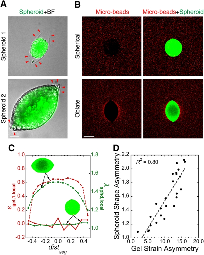
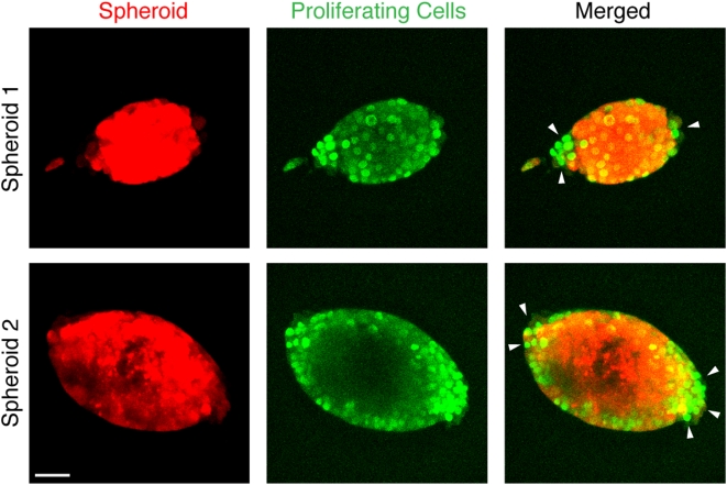
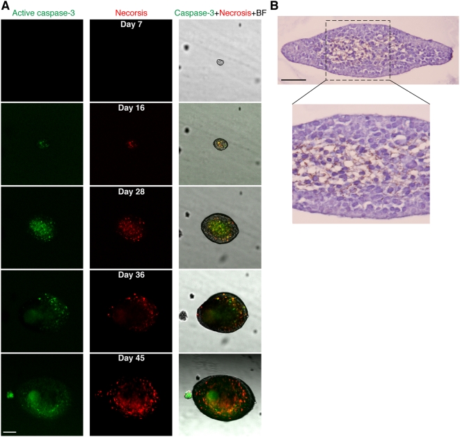
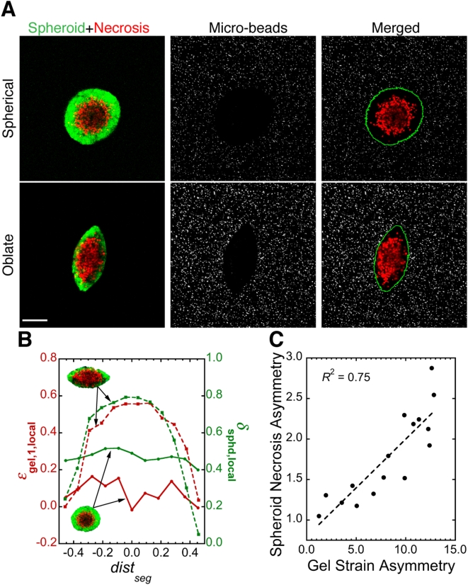
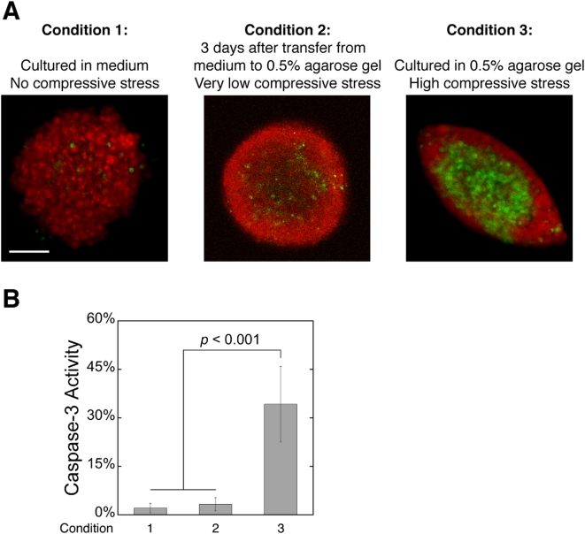
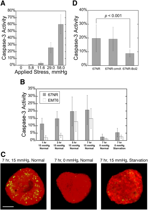
Methodology/principal findings: We co-embedded single cancer cells with fluorescent micro-beads in agarose gels and, using confocal microscopy, recorded the 3D distribution of micro-beads surrounding growing spheroids. The change in micro-bead density was then converted to strain in the gel, from which we estimated the spatial distribution of compressive stress around the spheroids. We found a strong correlation between the peri-spheroid solid stress distribution and spheroid shape, a result of the suppression of cell proliferation and induction of apoptotic cell death in regions of high mechanical stress. By compressing spheroids consisting of cancer cells overexpressing anti-apoptotic genes, we demonstrate that mechanical stress-induced apoptosis occurs via the mitochondrial pathway.

Conclusions/significance: Our results provide detailed, quantitative insight into the role of micro-environmental mechanical stress in tumor spheroid growth dynamics, and suggest how tumors grow in confined locations where the level of solid stress becomes high. An important implication is that apoptosis via the mitochondrial pathway, induced by compressive stress, may be involved in tumor dormancy, in which tumor growth is held in check by a balance of apoptosis and proliferation.


Conflict of interest statement

Competing Interests: The authors have declared that no competing interests exist.

Figures

Figure 1
Figure 1. Mechanical stress accumulates around growing tumor spheroids.
(A) A growing spheroid (green) and its surrounding micro-beads (red). Scale bar = 100 µm. (B) Quantification of relative micro-bead density (ρ bead) in 10-µm thick shells of agarose gel around the growing spheroid shown in A as a function of the distance of the shell from spheroid center (dist). (C) Correlation between spheroid diameter (Dsphd) and the strain in the first 10-µm thick shell of agarose gel (ε gel,1) around spheroids. R is the linear regression coefficient; slope of the regression line is significantly greater than zero (p<0.0001).
Figure 2
Figure 2. Mechanical stress distribution controls tumor spheroid shape.
(A) Agarose gel can fail under tension from growing tumor spheroids (green). Red arrowheads indicate the edge of planar cracks in the agarose gel (BF: bright-field image taken in Nomarski mode). Scale bar = 50 µm. (B) Spheroids (green) of different shapes and their surrounding stress fields visualized by micro-beads (red). Scale bar = 150 µm. (C) Relationship between local strain in agarose gel (ε gel,1,local) and local spheroid deformation (λ sphd,local) for the spheroids (green, inset) shown in A. dist seg is the distance of spheroid segments from spheroid center normalized over the length of the major axis. (D) Correlation between the asymmetry in spheroid shape and in the corresponding strain in the surrounding agarose gel, showing that spheroids are more deformed along the direction of higher stress. Each data point is for one spheroid. R is the linear regression coefficient; slope of the regression line is significantly greater than zero (p<0.0001). Methods for image analysis in C and D are described in Supplementary Methods S1, Supplementary Fig. S2, Movie S2 and Movie S3.
Figure 3
Figure 3. Cancer cell proliferation (green) in tumor spheroids (red) is suppressed in the direction of higher mechanical stress (i.e., in the direction of the minor axis of oblate spheroids).
Arrowheads indicate the regions with more cell proliferation. Scale bar = 50 µm.
Figure 4
Figure 4. Mechanical stress induces apoptotic cell death in tumor spheroids.
(A) The development of apoptosis (green) and secondary necrosis (red) in a growing spheroid embedded in 0.5% agarose gel. Scale bar = 100 µm. (B) Immunohistochemical staining (TUNEL and hematoxylin) confirming the results in A. Image in the lower panel shows detail of the area within the dashed line in the image in the upper panel. Scale bar = 50 µm.
Figure 5
Figure 5. Mechanical stress distribution correlates strongly with the distribution of cell death in tumor spheroids.
(A) Live spheroids (green) of different shapes, their internal cell death (red), and their surrounding stress fields visualized by micro-beads (gray). The green line in the right column shows the edge of spheroids. Scale bar = 100 µm. (B) Relationship between local strain in agarose gel (ε gel,1,local) and local necrotic fraction in spheroids (δ sphd,local) for the two spheroids shown in A. (C) Correlation between the asymmetry in spheroid necrotic fraction and in the corresponding strain in the surrounding agarose gel, showing that there is more cell death along the direction of higher compressive stress. Each data point is for one spheroid. R is the linear regression coefficient; slope of the regression line is significantly greater than zero (p<0.0001). Methods for image analysis in B and C are described in Supplementary Methods, Supplementary Fig. S2, Movie S3 and Movie S4.
Figure 6
Figure 6. Agarose gel toxicity or limitations of nutrients, growth factors and oxygen is not responsible for the apoptosis observed in spheroids under high levels of compressive stress.
(A) Caspase-3 activity (green) in tumor spheroids (red) cultured in free suspension (Condition 1), transferred to 0.5% agarose gel for 3 days after reaching plateau-phase in free suspension (Condition 2), or cultured from single cells in 0.5% agarose gel (Condition 3). Scale bar = 100 µm. (B) Quantification of the fraction of apoptotic cells in tumor spheroids cultured under the 3 conditions in A.
Figure 7
Figure 7. Mechanical stress-induced cell death in tumor spheroids acts via the mitochondrial pathway.
(A) Caspase-3 activity increases in monolayers of cancer cells in response to higher external stress. Cells were compressed for 17 hr. (B) Caspase-3 activity in spheroids made from two different cancer cell lines in response to different external stress (0 mmHg or 15 mmHg) and nutrient conditions (Normal: normal medium; Starvation: no glucose, no serum and 1% oxygen). (C) Typical caspase-3 activity (green) in spheroids (red) cultured in 3 of the conditions in B. Scale bar = 100 um. (D) Bcl-2 over-expression inhibits stress-induced apoptosis, but crmA transduction does not. Spheroids were compressed with 15 mmHg for 7 hr while being supplied with normal medium.

References

    1. Harris AL. Hypoxia–a key regulatory factor in tumour growth. Nat Rev Cancer. 2002;2:38–47. - PubMed
    1. Semenza GL. Targeting HIF-1 for cancer therapy. Nat Rev Cancer. 2003;3:721–732. - PubMed
    1. Folkman J. Tumor angiogenesis: therapeutic implications. N Engl J Med. 1971;285:1182–1186. - PubMed
    1. Carmeliet P, Jain RK. Angiogenesis in cancer and other diseases. Nature. 2000;407:249–257. - PubMed
    1. Jain RK. Normalization of tumor vasculature: an emerging concept in antiangiogenic therapy. Science. 2005;307:58–62. - PubMed

Publication types

LinkOut - more resources