. 2009;12(3):209-20.
doi: 10.1007/s10456-009-9139-3. Epub 2009 Feb 27.

Chloride intracellular channel 4 is involved in endothelial proliferation and morphogenesis in vitro

Affiliations

Chloride intracellular channel 4 is involved in endothelial proliferation and morphogenesis in vitro

Jennifer J Tung et al. Angiogenesis. 2009.

Abstract

New capillaries are formed through angiogenesis and an integral step in this process is endothelial tubulogenesis. The molecular mechanisms driving tube formation during angiogenesis are not yet delineated. Recently, the chloride intracellular channel 4 (CLIC4)-orthologue EXC-4 was found to be necessary for proper development and maintenance of the Caenorhabditis elegans excretory canal, implicating CLIC4 as a regulator of tubulogenesis. Here, we studied the role of CLIC4 in angiogenesis and endothelial tubulogenesis. We report the effects of inhibiting or inducing CLIC4 expression on distinct aspects of endothelial cell behavior in vitro. Our experiments utilized RNA interference to establish cultured human endothelial cell lines with significant reduction of CLIC4 expression, and a CLIC4-expressing lentiviral plasmid was used to establish CLIC4 overexpression in endothelial cells. We observed no effect on cell migration and a modest effect on cell survival. Reduced CLIC4 expression decreased cell proliferation, capillary network formation, capillary-like sprouting, and lumen formation. This suggests that normal endogenous CLIC4 expression is required for angiogenesis and tubulogenesis. Accordingly, increased CLIC4 expression promoted proliferation, network formation, capillary-like sprouting, and lumen formation. We conclude that CLIC4 functions to promote endothelial cell proliferation and to regulate endothelial morphogenesis, and is thus involved in multiple steps of in vitro angiogenesis.


Figures

Fig. 1
Fig. 1
Establishing CLIC4 knockdown and overexpression in endothelial cells. a Immunoblotting with polyclonal rabbit anti-CLIC4 antibody confirms knockdown of CLIC4 protein expression in shRNA3 and shRNA5 endothelial cell lines compared with plko sc control line. Immunoblotting with anti-CLIC1 antibody confirms that knockdown by shRNA is specific to CLIC4 and does not interfere with CLIC1 expression. b Immunoblotting with the anti-CLIC4 antibody confirms CLIC4 overexpression in the CLIC4 endothelial cell line compared with pccl control. Alpha-tubulin served as a loading control for all immunoblots. c Established HUVEC shRNA3 knockdown cells exhibited no major change in endothelial cell morphology when compared with plko sc control cells. HUVEC shRNA5 appear to be more rounded when compared with plko sc. HUVEC refer to endothelial cells that have not been infected with any constructs. d No major change was observed in endothelial cell morphology between pccl control cells and CLIC4-overexpressing cells
Fig. 2
Fig. 2
CLIC4 expression has no effect on endothelial cell migration. a CLIC4 knockdown cell lines shRNA3 and shRNA5 exhibited no noticeable change in directed endothelial cell migration 6 h after scraping in a migration assay when compared with control plko sc. b Likewise, CLIC4-overexpressing cells exhibited no noticeable change in endothelial cell migration 6 h after scraping when compared with control pccl
Fig. 3
Fig. 3
CLIC4 expression promotes endothelial cell proliferation. a Inhibition of CLIC4 expression by shRNA resulted in decreased endothelial cell proliferation when compared with plko sc control. b CLIC4 overexpression resulted in a significant increase in endothelial cell proliferation when compared with control pccl. (*P < 0.05; **P < 0.01)
Fig. 4
Fig. 4
Altering CLIC4 expression affects endothelial cell survival. a CLIC4 knockdown lines significantly increased cell survival at 48 h, but not 24 h post-seeding. b CLIC4 overexpression resulted in significantly decreased endothelial cell survival at 48 h, but not 24 h post-seeding. (*P < 0.05; **P < 0.01)
Fig. 5
Fig. 5
CLIC4 expression promotes in vitro endothelial network formation. a Microscopy of CLIC4 knockdown or overexpressing cells in a collagen network formation assay shows decreased network formation with CLIC4 knockdowns, while CLIC4 overexpression resulted in increased network formation when compared with respective controls. b Quantification confirms that CLIC4 knockdown results in decreased network formation by showing that reduced CLIC4 expression leads to a significant reduction in surface area coverage by endothelial cells. CLIC4 overexpression is shown to lead to increased network formation with a significant increase in surface area occupied by endothelial cells. Control standards were set at 100% surface area occupation. c Quantification of branchpoints for each knockdown cell line further confirms CLIC4 knockdown inhibition on endothelial network formation. Knockdown cell lines form significantly fewer branchpoints than control plko sc. Quantification was done by counting visible branchpoints per field of view at 10× objective. d CLIC4-overexpressing cells form significantly more branchpoints than control pccl. Quantification was done by counting visible branchpoints per field of view at 5× objective. One branchpoint was considered any focal intersection of two or more cords. (*P < 0.05; **P < 0.01)
Fig. 6
Fig. 6
Reduced CLIC4 expression inhibits endothelial network formation independent of its proliferative defect. a Comparison of network formation by knockdown cell lines and control cells treated with mitomycin C indicates that there is inhibition of network formation independent from reduced CLIC4 inhibition of proliferation. b A proliferation assay confirms mitomycin C growth arrest of plko sc control cells. c Quantification of surface area occupied by endothelial cells confirms an additional defect associated with reduced CLIC4 expression independent of reduced CLIC4 proliferation inhibition. Mitomycin C-treated control standards were set at 100% surface area occupation. d Quantification of branchpoints further confirms an additional defect associated with reduced CLIC4 expression separate from reduced CLIC4 proliferation inhibition. (*P < 0.05 compared with plko sc; **P < 0.01 compared with plko sc; +P < 0.05 compared with plko sc + mmc; ++P < 0.01 compared with plko sc + mmc)
Fig. 7
Fig. 7
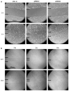
CLIC4 expression affects capillary-like sprouting in vitro. a Microscopy indicates that HUVEC shRNA3 have altered tube morphogenesis in that they tend to form shorter, more stunted tube structures as indicated by a red arrow. HUVEC shRNA5 show a decrease in the number of branches per sprout. HUVEC shRNA5 also exhibited stunted sprouts indicated by a red arrow. Unlike HUVEC shRNA3 cells, HUVEC shRNA5 cells did not form lumens. Black arrows indicate robust, lumen-containing sprouts. b Microscopy from a separate trial confirms the phenotypes of HUVEC shRNA3 stunted tube structures and HUVEC shRNA5 stunted sprouts without lumens. c Quantification of the number of lumen-containing sprouts shows that HUVEC shRNA5 exhibits a significant decrease in the number of lumen-containing sprouts formed when compared with plko sc control. d Microscopy of CLIC4-overexpressing endothelial cells show increased sprouting and lumen formation when compared with plko sc control. Lumen-containing sprouts are indicated by black arrows. e Quantification for CLIC4-overexpressing endothelial cells indicate a significant increase in the number of lumen-containing sprouts. (*P < 0.05; **P < 0.01)

Similar articles

Cited by

References

    1. Risau W. Mechanisms of angiogenesis. Nature. 1997;386:671–674. doi: 10.1038/386671a0. - DOI - PubMed
    1. Kamei M, Saunders WB, Bayless KJ, Dye L, Davis GE, Wein-stein BM. Endothelial tubes assemble from intracellular vacuoles in vivo. Nature. 2006;442:453–456. doi: 10.1038/nature04923. - DOI - PubMed
    1. Hogan BL, Kolodziej PA. Organogenesis: molecular mechanisms of tubulogenesis. Nat Rev Genet. 2002;3:513–523. doi: 10.1038/nrg840. - DOI - PubMed
    1. Berry KL, Bulow HE, Hall DH, Hobert O. A C. elegans CLIC-like protein required for intracellular tube formation and maintenance. Science. 2003;302:2134–2137. doi: 10.1126/science.1087667. - DOI - PubMed
    1. Ulmasov B, Bruno J, Woost PG, Edwards JC. Tissue and subcellular distribution of CLIC1. BMC Cell Biol. 2007;8:8. doi: 10.1186/1471-2121-8-8. - DOI - PMC - PubMed

Publication types

MeSH terms