Neural activation of swallowing and swallowing-related tasks in healthy young adults: an attempt to separate the components of deglutition
- PMID: 19247994
- PMCID: PMC6870848
- DOI: 10.1002/hbm.20743
Neural activation of swallowing and swallowing-related tasks in healthy young adults: an attempt to separate the components of deglutition
Abstract
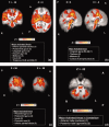
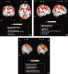
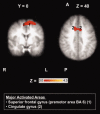
Understanding the underlying neural pathways that govern the highly complex neuromuscular action of swallowing is considered crucial in the process of correctly identifying and treating swallowing disorders. The aim of the present investigation was to identify the neural activations of the different components of deglutition in healthy young adults using functional magnetic resonance imaging (fMRI). Ten right-handed young healthy individuals were scanned in a 3-Tesla Siemens Allegra MRI scanner. Participants were visually cued for both a "Swallow" task and for component/control tasks ("Prepare to swallow", "Tap your tongue", and "Clear your throat") in a randomized order (event-related design). Behavioral interleaved gradient (BIG) methodology was used to address movement-related artifacts. Areas activated during each of the three component tasks enabled a partial differentiation of the neural localization for various components of the swallow. Areas that were more activated during throat clearing than other components included the posterior insula and small portions of the post- and pre-central gyri bilaterally. Tongue tapping showed higher activation in portions of the primary sensorimotor and premotor cortices and the parietal lobules. Planning did not show any areas that were more activated than in the other component tasks. When swallowing was compared with all other tasks, there was significantly more activation in the cerebellum, thalamus, cingulate gyrus, and all areas of the primary sensorimotor cortex bilaterally.
Figures


References
-
- Addington WR,Stephens RE,Gilliland KA ( 1999): Assessing the laryngeal cough reflex and the risk of developing pneumonia after stroke: An interhospital comparison. Stroke 30: 1203–1207. - PubMed
-
- Alberts MJ,Horner J,Gray L,Brazer SR ( 1992): Aspiration after stroke: Lesion analysis by brain MRI. Dysphagia 7: 170–173. - PubMed
-
- Andersen RA,Buneo CA ( 2002): Intentional maps in posterior parietal cortex. Annu Rev Neurosci 25: 189–220. - PubMed
-
- Augustine JR ( 1996): Circuitry and functional aspects of the insular lobe in primates including humans. Brain Res Rev 22: 229–244. - PubMed
-
- Barkmeier JM,Bielamowicz S,Takeda N,Ludlow CL ( 2002): Laryngeal muscle activation patterns during upright and supine swallowing. J Appl Physiol 93: 740–745. - PubMed
Publication types
MeSH terms
Substances
LinkOut - more resources
Full Text Sources
Miscellaneous

