

Editorial
doi: 10.1002/uog.6336.
Sonographic evaluation in the second stage of labor to improve the assessment of labor progress and its outcome
- PMID: 19247999
- PMCID: PMC3138397
- DOI: 10.1002/uog.6336
Item in Clipboard
Editorial
Sonographic evaluation in the second stage of labor to improve the assessment of labor progress and its outcome
Ultrasound Obstet Gynecol.
2009 Mar.
No abstract available
Figures

Partogram developed by Emanuel Friedman which describes the relationship between time in the horizontal axis and two variables: cervical dilatation and station of the presenting part. Reproduced from: Friedman EA. Dysfunctional labor. In Management of Labor, CohenWR, Friedman EA (eds). University Park Press: Baltimore, 1983: 11-2415.

“Angle of progression”. This is the angle formed between a line placed through the midline of the symphysis pubis, and a second line (in red) drawn from the inferior apex of the symphysis tangentially to the fetal skull. In this figure, the angle of progression is greater than 120 degrees. The original figure was provided by Professor Karim Kalache (Berlin, Germany) and has been slightly modified.

The relationship between the bony pelvis and the presenting part to define station. It is generally held that when the largest diameter of the fetal head is at the level of the pelvic inlet, the leading part of the head will be at the level of the ischial spines (station 0). The plus and minus numbers represent centimeters below and above the ischial spines, respectively. Reproduced from: Greenhill JP, Friedman EA. Biological Principles and Modern Practice of Obstetrics. WB Saunders: Philadelphia, 1974 40.

“Head progression distance”. This diagram describes the parameter originally proposed by Professor Hans Peter Dietz (Sydney, Australia) to assess the degree of descent of the presenting part. A line through the inferoposterior symphyseal margin (parallel to the main transducer axis) is the reference line. The minimal distance between this line and the leading edge of the presenting part is measured (line marked by calipers and is in red) and represents the head progression distance.

A diagram of the birth canal demonstrating that it has a curved trajectory. The presenting part descends in a straight line, and then curves as it approaches the outlet. This figure is reproduced with kind permission from Greenhill JP and Friedman EA, Biological Principles and Modern Practice of Obstetrics; W.B. Saunders Company, 197440.
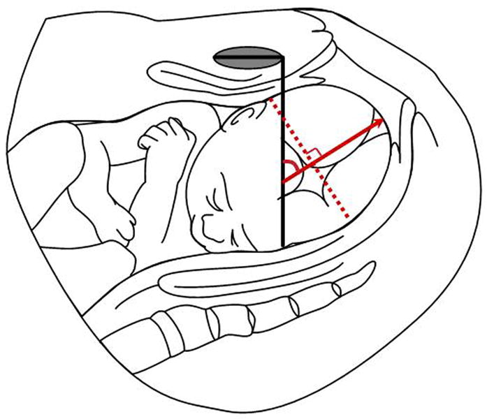
“Head direction” as described by Professor Wolfgang Henrich et al (Berlin, Germany). An infrapubic line (in black) is drawn perpendicular to the long axis of the symphysis pubis. The red dashed line represents the widest fetal head diameter. The head direction is the angle formed between the infrapubic line and another line (red arrow line) drawn perpendicular to the widest diameter of the fetal head. When this line (red arrow line) points ventrally at an angle of 30 degrees or more, it is considered “head up.” For lines below 0 degrees, the direction is called “head down,” and all other angles are considered horizontal.
Comment in
-
What is the best transperineal ultrasound parameter for predicting success of vacuum extraction?Ultrasound Obstet Gynecol. 2009 Jun;33(6):735; author reply 736. doi: 10.1002/uog.6400. Ultrasound Obstet Gynecol. 2009. PMID: 19434619 No abstract available.
References
-
- Dietz HP, Lanzarone V, Simpson JM. Predicting operative delivery. Ultrasound Obstet Gynecol. 2006;27:409–15. - PubMed
-
- Calkins LA. On predicting the length of labor: I. First stage. Am J Obstet Gynecol. 1941;42:802–11.
-
- Calkins LA. Second stage of labor: The descent phase. Am J Obstet Gynecol. 1944;48:798–803.
-
- Friedman EA. The graphic analysis of labor. Am J Obstet Gynecol. 1954;68:1568–75. - PubMed
-
- Friedman EA. Cervimetry: an objective method for the study of cervical dilatation in labor. Am J Obstet Gynecol. 1956;71:1189–93. - PubMed
Publication types
MeSH terms
Grants and funding
LinkOut - more resources
Full Text Sources
