

Nodular liver lesions involving multiple myeloma: a case report and literature review
- PMID: 19248205
- PMCID: PMC2653410
- DOI: 10.3748/wjg.15.1014
Nodular liver lesions involving multiple myeloma: a case report and literature review
Abstract
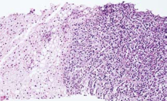
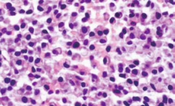
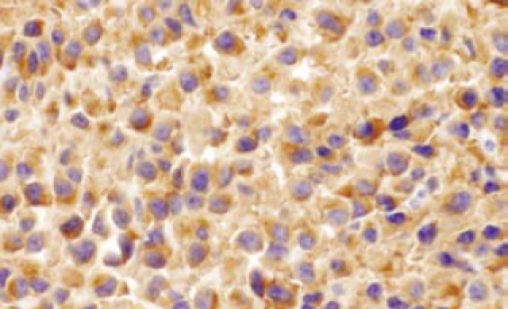
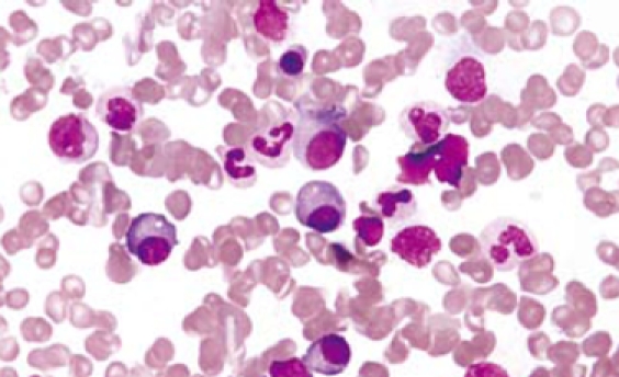
We report a case of a 62-year old woman admitted to our hospital for multiple nodular metastatic liver lesions found by ultrasonography in a regular medical examination. Routine laboratory tests were normal. PET-CT showed multiple bone lesions and nodular liver lesions. Liver biopsy revealed nodular infiltration of multiple myeloma with positive staining of kappa light chain. Further investigation of bone marrow aspiration, immunofixation and immunoelectrophoresis of serum protein, urine test for Bence-Jones protein, beta(2)-microglobulin in serum and urine confirmed the diagnosis. The patient also coinfected with hepatitis C virus (HCV). With six cycles of chemotherapy with VAD schedule, she achieved complete remission. In this report, a literature review of liver lesions involving multiple myeloma is also provided.
Figures

References
-
- Thomas FB, Clausen KP, Greenberger NJ. Liver disease in multiple myeloma. Arch Intern Med. 1973;132:195–202. - PubMed
-
- Perez-Soler R, Esteban R, Allende E, Tornos Salomo C, Julia A, Guardia J. Liver involvement in multiple myeloma. Am J Hematol. 1985;20:25–29. - PubMed
-
- Talamo G, Cavallo F, Zangari M, Barlogie B, Lee CK, Pineda-Roman M, Kiwan E, Krishna S, Tricot G. Clinical and biological features of multiple myeloma involving the gastrointestinal system. Haematologica. 2006;91:964–967. - PubMed
-
- Oshima K, Kanda Y, Nannya Y, Kaneko M, Hamaki T, Suguro M, Yamamoto R, Chizuka A, Matsuyama T, Takezako N, et al. Clinical and pathologic findings in 52 consecutively autopsied cases with multiple myeloma. Am J Hematol. 2001;67:1–5. - PubMed
-
- Thiruvengadam R, Penetrante RB, Goolsby HJ, Silk YN, Bernstein ZP. Multiple myeloma presenting as space-occupying lesions of the liver. Cancer. 1990;65:2784–2786. - PubMed
Publication types
MeSH terms
Substances
LinkOut - more resources
Full Text Sources
Medical
Research Materials
