. 2009 Mar 6;136(5):876-90.
doi: 10.1016/j.cell.2009.02.014. Epub 2009 Feb 26.

STIM1 clusters and activates CRAC channels via direct binding of a cytosolic domain to Orai1

Affiliations

STIM1 clusters and activates CRAC channels via direct binding of a cytosolic domain to Orai1

Chan Young Park et al. Cell. .

Abstract

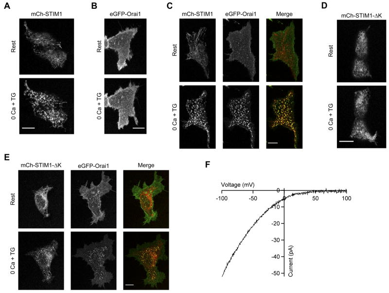
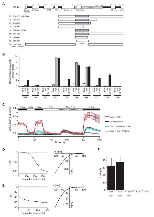
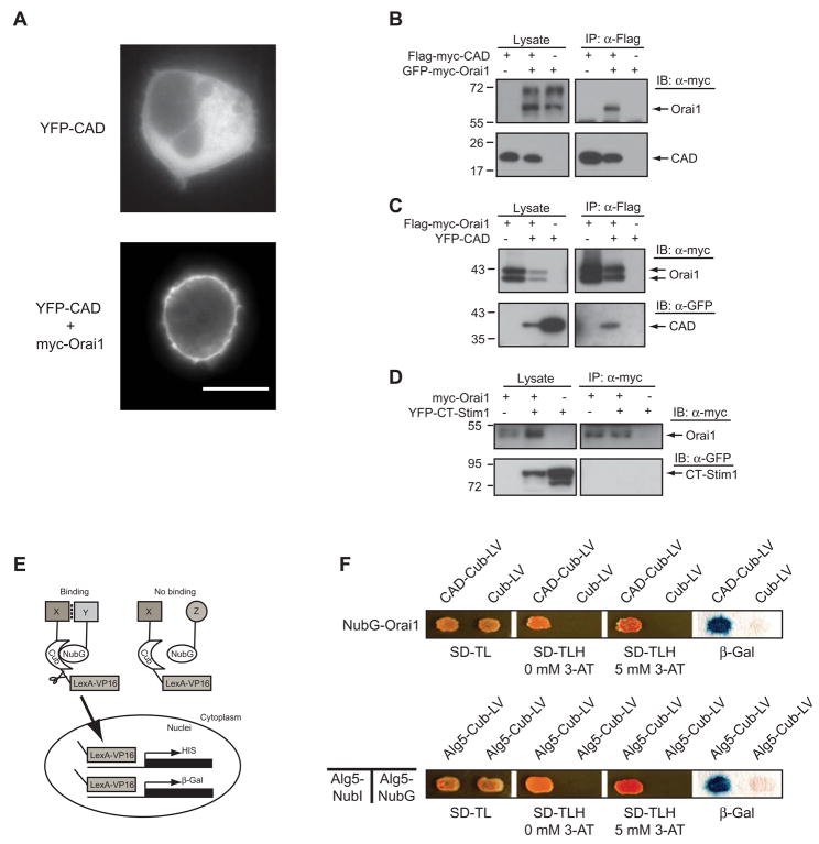
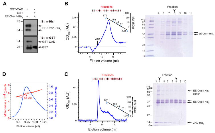
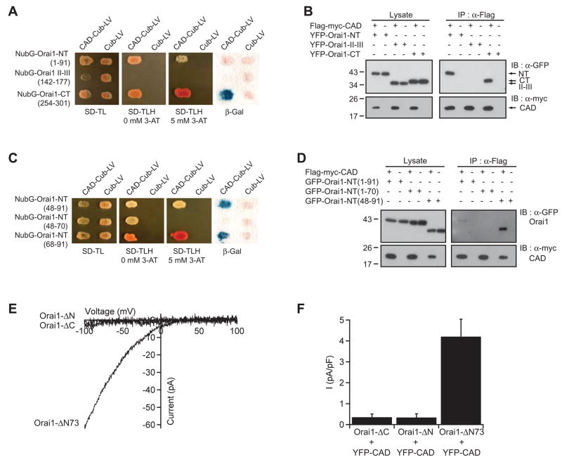
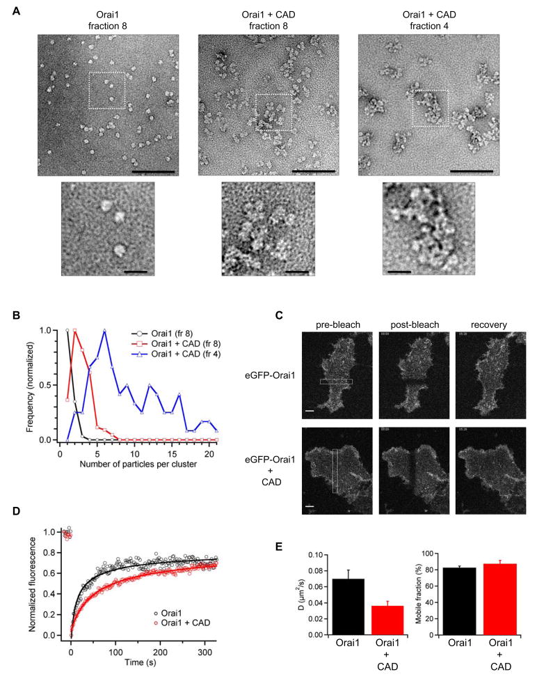
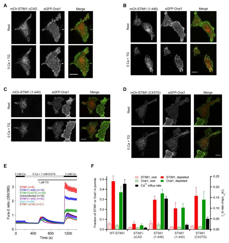
Store-operated Ca(2+) channels activated by the depletion of Ca(2+) from the endoplasmic reticulum (ER) are a major Ca(2+) entry pathway in nonexcitable cells and are essential for T cell activation and adaptive immunity. After store depletion, the ER Ca(2+) sensor STIM1 and the CRAC channel protein Orai1 redistribute to ER-plasma membrane (PM) junctions, but the fundamental issue of how STIM1 activates the CRAC channel at these sites is unresolved. Here, we identify a minimal, highly conserved 107-aa CRAC activation domain (CAD) of STIM1 that binds directly to the N and C termini of Orai1 to open the CRAC channel. Purified CAD forms a tetramer that clusters CRAC channels, but analysis of STIM1 mutants reveals that channel clustering is not sufficient for channel activation. These studies establish a molecular mechanism for store-operated Ca(2+) entry in which the direct binding of STIM1 to Orai1 drives the accumulation and the activation of CRAC channels at ER-PM junctions.


Figures

Figure 1
Figure 1. Deletion of the STIM1 Polybasic Domain Distinguishes Orai1-dependent and independent Mechanisms of STIM1 Accumulation at ER-PM Junctions
(A) Wild-type mCh-STIM1 expressed alone in HEK 293 cells redistributes from a diffuse ER distribution (Rest) to the cell periphery after store depletion with TG (0 Ca + TG). After depletion, puncta are visible at the cell footprint. (B) eGFP-myc-Orai1 when expressed alone does not redistribute into puncta after depletion. (C) When expressed together, STIM1 and Orai1 form colocalized puncta after store depletion. (D) Unlike wild-type STIM1, STIM1-ΔK expressed alone fails to form puncta; however, when coexpressed with Orai1 both proteins form colocalized puncta after store depletion (E). (F) Store depletion activates ICRAC in HEK 293 cells expressing STIM1-ΔK and Orai1 demonstrating that the polybasic region is not essential for ICRAC activation. All images were taken of cell footprints using confocal microscopy. Scale bars, 10 μm.
Figure 2
Figure 2. Identification of CAD as a Potent CRAC Channel Activator
(A) Full-length STIM1 with its putative functional domains (top) and truncated versions of STIM1 (D1-D9; bottom) with the CAD shown in gray. (B) NFAT-dependent luciferase activity in HEK 293T (NFAT-Luc) cells transfected with wild-type (WT) or truncated (D1-D9) STIM1 constructs. Cells were treated with 1 μM PMA or 1 μM TG+PMA as shown. D5 (CAD) is the minimal region that is necessary and sufficient to activate NFAT. Data are shown as mean ± sem (n=4). (C) CAD activates SOCE without depleting intracellular Ca2+ stores. Mean [Ca2+]i (± sem) in untransfected HEK 293 cells (black; n=28) and cells expressing CAD + Orai1 (red; n=15), CAD + Orai1E106A (gray; n=17), or CAD (342–440) + Orai1 (blue; n=12). (D) (left) ICRAC develops slowly in a representative cell cotransfected with WT GFP-STIM1 + myc-Orai1. Current recorded during brief pulses to −100 mV in 2 mM Ca2+o is plotted against time after break-in. (right) Characteristic I–V relationship for ICRAC recorded in 20 mM Ca2+o from the same cell. (E) (left) ICRAC is constitutively active in a representative cell cotransfected with YFP-CAD + myc-Orai1. Current is plotted as in D. (right) I–V relationship for the CAD-induced current recorded in 20 mM Ca2+o from the same cell. (F) Current densities in cells transfected with CAD or Orai1 alone, or cotransfected with myc-Orai1 and GFP-STIM1, YFP-CAD, or YFP-CT-STIM1. Current measured at −100 mV during voltage ramps in 20 mM Ca2+o was normalized to the cell capacitance. Mean values ± sem from 4–5 cells are shown.
Figure 3
Figure 3. CAD Associates with Orai1
(A) YFP-CAD is cytosolic when expressed by itself in a HEK 293 cell (top), but accumulates at the cell perimeter when coexpressed with Orai1 (bottom). Scale bar, 10 μm. (B, C) Western blots of cell lysates (left) or immunoprecipitated material (right) from cells expressing CAD, Orai1, or CAD + Orai1. Anti-Flag antibodies co-immunoprecipitate GFP-myc-Orai1 with Flag-myc-CAD (B), and co-immunoprecipitate YFP-CAD with Flag-myc-Orai1 (C). (D) CT-STIM1 does not co-immunoprecipitate with myc-Orai1 (representative of 4 experiments). (E) Schematic depiction of the yeast split ubiquitin assay. (F) CAD associates with Orai1 in the split ubiquitin assay. Yeast containing NubG-Orai1 and either Cub-LV alone or CAD-Cub-LV, grow well on plates lacking tryptophan and leucine but containing histidine (SD-TL). Only yeast expressing NubG-Orai1 and CAD-Cub-LV grow on plates lacking all three amino acids (SD-TLH) in the absence or presence of 5 mM 3-aminotriazole (3AT), a competitive HIS3 inhibitor that increases the stringency of selection. Yeast containing CAD-Cub-LV and NubG-Orai1 also activate LacZ whereas cells expressing NubG-Orai1 and Cub do not. The homomeric interaction of Alg5 is shown as a positive control.
Figure 4
Figure 4. CAD Binds Directly to Orai1
(A) Glutathione beads precipitate EE-Orai1-His8 with GST-CAD but not GST alone. (B) (Left) Size-exclusion profile of purified EE-Orai1-His8. On SDS-PAGE (right), peak fractions contain EE-Orai1-His8 as a pair of glycosylated and unglycosylated bands at ~37 kDa. (C) (Left) Size-exclusion profile of EE-Orai1-His8 and CAD-His6 coexpressed in Hi5 cells and affinity purified in 0.5 M NaCl. Much of the CAD and Orai1 co-elute in the void volume (MW > 10 MDa) of the Superose 6 gel filtration column (fraction 4). The band corresponding to the dimer of EE-Orai1-His8 (arrow) was identified by Western blotting. (D) Multi-angle light scattering (MALS) indicates a molecular weight of ~58 kDa for purified CAD-His6, approximately four times the predicted mass of the monomer.
Figure 5
Figure 5. CAD Binds to Both the N- and C-termini of Orai1
(A) In this split ubiquitin assay,β-gal production and growth of transformants on plates lacking histidine indicate a strong interaction between CAD and the C-terminus of Orai1, a weaker interaction with the N-terminus, and lack of interaction with the II–III loop of Orai1. (B) CAD in HEK 293T cells co-immunoprecipitates with YFP-tagged N-terminal and C-terminal fragments of Orai1 but not with the II–III loop. (C) Split ubiquitin assays showing that CAD interacts with aa 48–91 of the Orai1 N-terminus; this appears to be due to binding to aa 68–91 rather than aa 48–70. (D) CAD co-immunoprecipitates with Orai1-NT(48–91) but only very weakly with the entire N-terminus of Orai1 (aa 1–91). (E) Whole-cell recordings in 20 mM Ca2+o from HEK 293 cells co-expressing truncated Orai1-GFP proteins and YFP-CAD. Deletion of the N- or C-terminus of Orai1 abrogates function, but a substantial level of ICRAC is generated by Orai1-ΔN73. (F) Summary of ICRAC measurements (current at −100 mV, 20 mM Ca2+o, normalized to cell capacitance) with the indicated constructs (4 cells each, mean ± sem).
Figure 6
Figure 6. CAD Links Multiple CRAC Channels to Form Clusters
(A) Negative stain electron microscopy of purified EE-Orai1-His8 (left; fraction 8 from Fig. 4B), or complexes of EE-Orai1-His8 and CAD-His6 (right panels; fractions 4 and 8 from Fig. 4C). Scale bars, 100 nm (top), 20 nm (bottom, enlargements from dashed boxes). (B) Quantitation of cluster sizes for each condition shown in A; Orai1 (n=174), Orai1 + CAD (fr 8, n=134; fr 4, n=90). Each histogram is normalized to its maximum bin value. (C) FRAP of HEK 293 cells expressing eGFP-Orai1 (top row) or eGFP-Orai1 + CAD (bottom row). Images are shown at the indicated times after bleaching a bar across the cell footprint (left). Scale bars, 5 μm. (D) Time course of FRAP from single cells expressing eGFP-Orai1 alone (black) or with CAD (red). The superimposed fits indicate diffusion coefficients of 0.12 μm2/s (Orai1) and 0.026 μm2/s (Orai1 + CAD). (E) Mean diffusion coefficients and mobile fractions of GFP-Orai1 expressed alone (n=9) or with CAD (n=6; means ± sem).
Figure 7
Figure 7. CRAC Channel Clustering and Activation by STIM1 are Separable and Require the CAD Region
(A) When coexpressed with Orai1, STIM1-ΔCAD fails to form puncta and cluster Orai1 after store depletion. In contrast, STIM11–448 (B), STIM11–440 (C), and STIM1 C437G (D) co-accumulate with Orai1 in puncta after store depletion. In A–D, images are confocal micrographs of the HEK 293 cell footprint. Scale bars, 10 μm. (E) Ca2+ measurements (mean ± sem) in HEK 293 cells expressing the indicated constructs. In all cases, Orai1 was tagged with eGFP and the STIM1 or its variant was tagged with mCherry. (F) Puncta formation and Ca2+ influx in cells expressing Orai1 and STIM1 variants. Each set of bars shows the fraction of the cell s STIM1 or Orai1 fluorescence that is colocalized to puncta before and after store depletion (mean ± sem, n=4 cells for each) and the initial rate of Ca2+ entry (dR/dt, where R is the fura-2 350/380 fluorescence ratio) measured 10–20 s after readdition of Ca2+ in E.

Comment in

References

    1. Baba Y, Hayashi K, Fujii Y, Mizushima A, Watarai H, Wakamori M, Numaga T, Mori Y, Iino M, Hikida M, Kurosaki T. Coupling of STIM1 to store-operated Ca2+ entry through its constitutive and inducible movement in the endoplasmic reticulum. Proc Natl Acad Sci U S A. 2006;103:16704–16709. - PMC - PubMed
    1. Bautista DM, Hoth M, Lewis RS. Enhancement of calcium signalling dynamics and stability by delayed modulation of the plasma-membrane calcium-ATPase in human T cells. J Physiol. 2002;541:877–894. - PMC - PubMed
    1. Berridge MJ. Capacitative calcium entry. Biochem J. 1995;312:1–11. - PMC - PubMed
    1. Bolotina VM. Orai, STIM1 and iPLA2beta: a view from a different perspective. J Physiol. 2008;586:3035–3042. - PMC - PubMed
    1. Brandman O, Liou J, Park WS, Meyer T. STIM2 is a feedback regulator that stabilizes basal cytosolic and endoplasmic reticulum Ca2+ levels. Cell. 2007;131:1327–1339. - PMC - PubMed

Publication types