. 2009 Jun;21(6):996-1006.
doi: 10.1016/j.cellsig.2009.02.010. Epub 2009 Feb 25.

Differences in Galpha12- and Galpha13-mediated plasma membrane recruitment of p115-RhoGEF

Affiliations

Differences in Galpha12- and Galpha13-mediated plasma membrane recruitment of p115-RhoGEF

Raja Bhattacharyya et al. Cell Signal. 2009 Jun.

Abstract

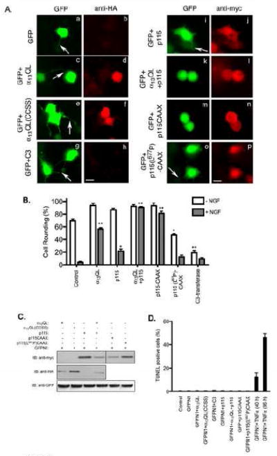
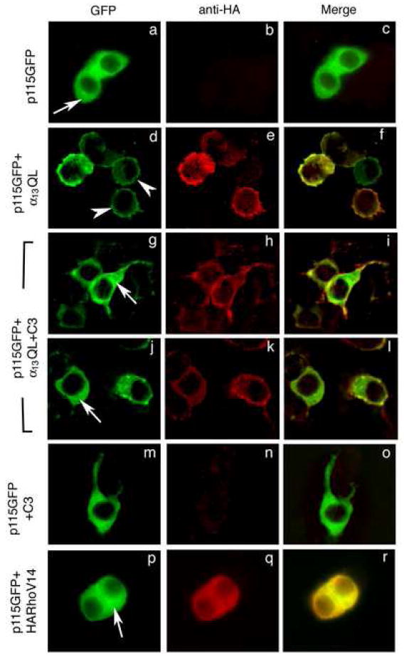
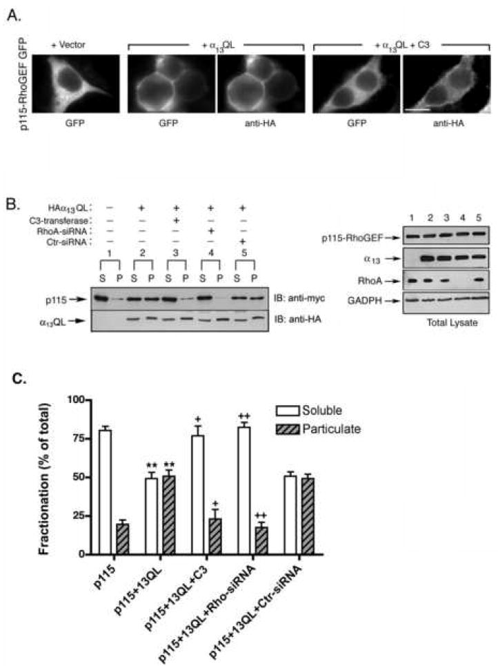
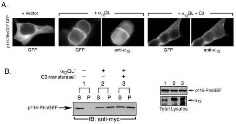
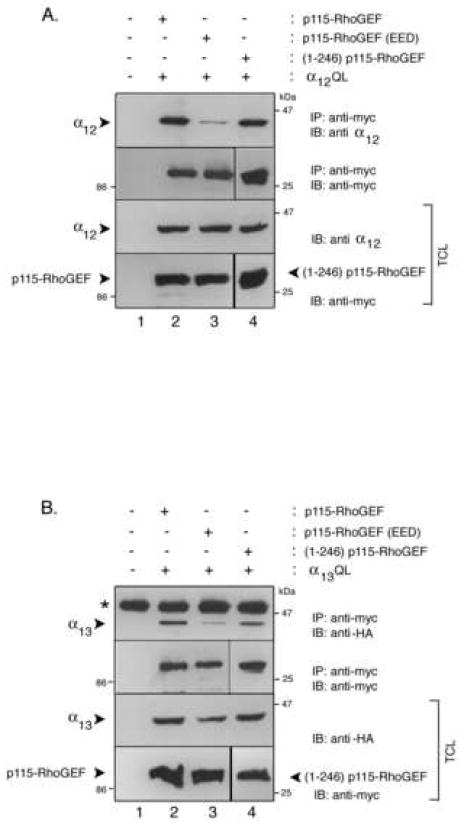
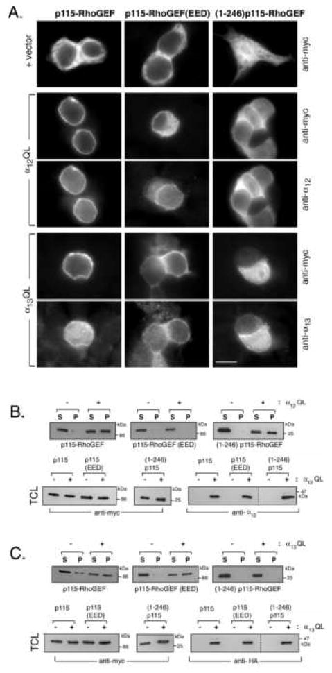
Regulator of G protein signaling domain-containing Rho guanine-nucleotide exchange factors (RGS-RhoGEFs) directly links activated forms of the G12 family of heterotrimeric G protein alpha subunits to the small GTPase Rho. Stimulation of G(12/13)-coupled GPCRs or expression of constitutively activated forms of alpha(12) and alpha(13) has been shown to induce the translocation of the RGS-RhoGEF, p115-RhoGEF, from the cytoplasm to the plasma membrane (PM). However, little is known regarding the functional importance and mechanisms of this regulated PM recruitment, and thus PM recruitment of p115-RhoGEF is the focus of this report. A constitutively PM-localized mutant of p115-RhoGEF shows a much greater activity compared to wild type p115-RhoGEF in promoting Rho-dependent neurite retraction of NGF-differentiated PC12 cells, providing the first evidence that PM localization can activate p115-RhoGEF signaling. Next, we uncovered the unexpected finding that Rho is required for alpha(13)-induced PM translocation of p115-RhoGEF. However, inhibition of Rho did not prevent alpha(12)-induced PM translocation of p115-RhoGEF. Additional differences between alpha(13) and alpha(12) in promoting PM recruitment of p115-RhoGEF were revealed by analyzing RGS domain mutants of p115-RhoGEF. Activated alpha(12) effectively recruits the isolated RGS domain of p115-RhoGEF to the PM, whereas alpha(13) only weakly does. On the other hand, alpha(13) strongly recruits to the PM a p115-RhoGEF mutant containing amino acid substitutions in an acidic region at the N-terminus of the RGS domain; however, alpha(12) is unable to recruit this p115-RhoGEF mutant to the PM. These studies provide new insight into the function and mechanisms of alpha(12/13)-mediated PM recruitment of p115-RhoGEF.


Figures

Figure 1
Figure 1. Rho-dependent neurite retraction in PC12 cells by overexpression of α13QL, p115-RhoGEF and their mutants
A. PC12 cells were transfected with plasmids encoding GFP along with vector (a and b), HA-α13QL (c and d), HA-α13QL(CCSS) (e and f), myc-tagged C3-transferase (g and h), myc-tagged p115-RhoGEF (i and j), myc-p115-RhoGEF and HA-α13QL (k and l), myc-p115-RhoGEF-CAAX (m and n), and myc-p115(L677P)-CAAX (o and p). 24 h post-transfection cells were transferred into serum free medium containing 100 ng/ml NGF. 16 h post-NGF-treatment cells were fixed and labeled with anti-HA antibody (anti-HA) or anti-myc antibody (anti-myc) followed by Texas Red-conjugated anti-mouse secondary antibody. The neuronal morphology was detected by observing the distribution of GFP in the cells co-expressing the indicated proteins. Representative images are presented. The arrows indicate neurite formation. Note that the GFP images are purposely overexposed so that the neurites are visible. Bar, 10 μm. B. A quantitative analysis of cell rounding is represented by plotting percent (%) of round cells calculated in each transfection experiment, as described in “Materials and Methods.” The data are average +/- S.E. of three separate experiments. Statistical significance (student’s t-test), indicated by * (p < 0.05) and ** (p < 0.001), is shown and compares untreated samples (-NGF) to control and NGF-treated (+NGF) to control. C. Total cell lysates from the indicated transfections of PC12 cells were immunoblotted with anti-myc, anti-HA and anti-GFP antibodies to determine expression levels of GFP, p115-RhoGEF, α13QL and their mutants. D. 40 h post-transfection, differentiating PC12 cells were subjected to TUNEL assay as described in the “Materials and Methods” section. The data is a representative experiment performed in triplicate.
Figure 2
Figure 2. Inhibition of Rho prevents α13QL-induced PM translocation of p115-RhoGEF in PC12 cells
PC12 cells were transiently transfected with plasmids encoding GFP-tagged p115-RhoGEF (p115GFP), C3-transferase (C3), HA-tagged RhoV14 (HARhoV14) or α13QL (α13QL), in various combinations as indicated. 16 h after transfection, cells were fixed and stained with anti-HA monoclonal antibody (b, e, h, k, n, and q) followed by anti-mouse secondary antibody conjugated to Texas Red to detect α13QL or RhoV14. p115-RhoGEF was detected due to the intrinsic fluorescence of the fused GFP (a, d, g, j, m, and p). Arrows indicate cytoplasmic staining, and arrowheads indicate plasma membrane staining. Representative images were recorded by immunofluorescence microscopy.
Figure 3
Figure 3. Inhibition or depletion of Rho prevents α13QL-induced PM recruitment of p115-RhoGEF in HEK293 cells
A. HEK293 cells were transfected with the expression vector for p115-RhoGEF-GFP along with empty vector, an expression plasmid for HA-tagged α13QL, or expression plasmids for both HA-tagged and α13QL myc-tagged C3-transferase (C3), as indicated. 24 h post-transfection, cells were fixed and subjected to immunofluorescence microscopy as described in the “Materials and Methods.” B. HEK293 cells were transfected with plasmids encoding myc-tagged p115-RhoGEF along with expression plasmids for HA-tagged α13QL, myc-tagged C3-transferase, siRNA specific for Rho silencing (RhoA-siRNA) or non-specific siRNA (Ctr-siRNA), as indicated (left panel). Cells were lysed and fractionated into soluble (S) and particulate (P) fractions, as described in the “Materials and Methods.” The fractions were immunoblotted (IB) with anti-myc antibody to detect p115-RhoGEF and anti-HA antibody to detect α13QL. Total cell lysates were immunoblotted (right panel) with anti-myc antibody and anti-HA antibody to show equivalent expression of p115-RhoGEF and α13QL, respectively. In addition, effective knockdown of endogenous Rho was detected by probing cell lysates with anti-Rho antibody, and immunoblotting with an anti-GADPH antibody served as a loading control. The lane numbering in the right panel indicates that the cell lysate corresponds to the transfection indicated in the left panel. C. Data from three independent fractionation experiments of p115-RhoGEF were quantitated (mean +/- S.E.). Statistical significance (student’s t-test), indicated by ** (p < 0.005), compares p115-RhoGEF alone to p115-RhoGEF in the presence of co-expressed α13QL; + (p < 0.05) and ++ (p < 0.005) indicates significance in comparison to p115-RhoGEF + α13QL. Soluble fractions (white bars) are compared to other soluble fractions, while particulate fractions (black bars) are compared to other particulate fractions.
Figure 4
Figure 4. α12QL-induced PM recruitment of p115-RhoGEF is refractory to inhibition of Rho
A. HEK293 cells were transfected with the expression vector for p115-RhoGEF-GFP along with empty vector, an expression plasmid for α12QL, or expression plasmids for both α12QL and myc-tagged C3-transferase (C3), as indicated. 24 h post-transfection, cells were fixed and subjected to immunofluorescence microscopy as described in the “Materials and Methods.” B. HEK293 cells were transfected with plasmids encoding myc-tagged p115-RhoGEF along with expression plasmids for α12QL and/or myc-tagged C3-transferase, as indicated (left panel). Cells were lysed and fractionated into soluble (S) and particulate (P) fractions, as described in the “Materials and Methods.” The fractions were immunoblotted (IB) with anti-myc antibody to detect p115-RhoGEF. Total cell lysates were immunoblotted (right panel) with anti-myc antibody and anti-α12 antibody to show expression of p115-RhoGEF and α12QL, respectively. The lane numbering in the right panel indicates that the cell lysate corresponds to the transfection indicated in the left panel.
Figure 5
Figure 5. Co-immunoprecipitation of α12QL and α13QL with p115-RhoGEF and p115-RhoGEF RGS domain mutants
A. HEK293 cells were transfected with empty vector (lane 1), expression plasmids for α12QL and p115-RhoGEF (lane 2), expression plasmids for α12QL and p115-RhoGEF(EED) (lane 3), or expression plasmids for α12QL and (1-246)p115-RhoGEF (lane 4). Cells were lysed and the lysates were subjected to immunoprecipitation (IP) by monoclonal anti-myc antibody to precipitate p115-RhoGEF and its mutants. The immunoprecipitates were immunoblotted (IB) with polyclonal anti-α12 antibody (upper panel) and anti-myc antibody (second panel). The corresponding total cell lysates were immunoblotted and probed with anti-myc or anti-α12 antibodies to show expression of p115-RhoGEF and its mutants (lower panel), and α12QL (third panel). B. HEK293 cells were transfected with empty vector (lane 1), expression plasmids for α13QL and p115-RhoGEF (lane 2), expression plasmids for α13QL and p115-RhoGEF(EED) (lane 3), or expression plasmids for α13QL and (1-246)p115-RhoGEF (lane 4). Cells were lysed and the lysates were subjected to immunoprecipitation (IP) by monoclonal anti-myc antibody to precipitate p115-RhoGEF and its mutants. The immunoprecipitates were immunoblotted (IB) with polyclonal anti-HA antibody (upper panel) to detect α13QL and anti-myc antibody (second panel). The corresponding total cell lysates were immunoblotted and probed with anti-myc or anti-HA antibodies to show expression of p115-RhoGEF and its mutants (lower panel), and α13QL (third panel).
Figure 6
Figure 6. Differential α12QL- and α13QL-mediated PM recruitment of p115-RhoGEF RGS domain mutants
A. HEK293 cells were transfected with either p115-RhoGEF (left column), p115-RhoGEF(EED) (middle column), or (1-246)p115-RhoGEF (right column). Each p115-RhoGEF wt or mutant was co-transfected with empty vector (upper row), α12QL (rows 2 and 3), or α13QL (rows 4 and 5). 24 h post-transfection, cells were fixed and subjected to immunofluorescence microscopy as described in the “Materials and Methods,” using the indicated antibodies. B. HEK293 cells were transfected with either p115-RhoGEF, p115-RhoGEF(EED), or (1-246)p115-RhoGEF. Each p115-RhoGEF wt or mutant was co-transfected with empty vector or α12QL, as indicated. Cells were lysed and fractionated into soluble (S) and particulate (P) fractions, as described in the “Materials and Methods.” The fractions were immunoblotted (IB) with anti-myc antibody to detect p115-RhoGEF (upper panels). Total cell lysates (TCL) were immunoblotted with anti-myc antibody and anti-α12 antibody to show expression of p115-RhoGEF and α12QL, respectively (lower panels). C. HEK293 cells were transfected with either p115-RhoGEF, p115-RhoGEF(EED), or (1-246)p115-RhoGEF. Each p115-RhoGEF wt or mutant was co-transfected with empty vector or α13QL, as indicated. Cells were lysed and fractionated into soluble (S) and particulate (P) fractions, as described in the “Materials and Methods.” The fractions were immunoblotted (IB) with anti-myc antibody to detect p115-RhoGEF (upper panels). Total cell lysates were immunoblotted with anti-myc antibody and anti-HA antibody to show expression of p115-RhoGEF and α13QL, respectively (lower panels). The dashed lines (lower right panels in B and C) indicate that the samples are from the same gel; intervening lanes that were irrelevant to the figure were simply removed.

References

    1. Cabrera-Vera TM, Vanhauwe J, Thomas TO, Medkova M, Preininger A, Mazzoni MR, Hamm HE. Endocr Rev. 2003;24(6):765–781. - PubMed
    1. Kelly P, Casey PJ, Meigs TE. Biochemistry. 2007;46(23):6677–6687. - PubMed
    1. Rossman KL, Der CJ, Sondek J. Nat Rev Mol Cell Biol. 2005;6(2):167–180. - PubMed
    1. Sternweis PC, Carter AM, Chen Z, Danesh SM, Hsiung YF, Singer WD. Adv Protein Chem. 2007;74:189–228. - PubMed
    1. Wells CD, Liu MY, Jackson M, Gutowski S, Sternweis PM, Rothstein JD, Kozasa T, Sternweis PC. J Biol Chem. 2002;277(2):1174–1181. - PubMed

Publication types

MeSH terms

Substances

LinkOut - more resources