

Review
doi: 10.1016/j.cellsig.2009.02.012.
Epub 2009 Feb 26.
PTH and PTHrP signaling in osteoblasts
Affiliations
- PMID: 19249350
- PMCID: PMC2723940
- DOI: 10.1016/j.cellsig.2009.02.012
Item in Clipboard
Review
PTH and PTHrP signaling in osteoblasts
Cell Signal.
2009 Aug.
Abstract
The striking clinical benefit of PTH in osteoporosis began a new era of skeletal anabolic agents. Several studies have been performed, new studies are emerging out and yet controversies remain on PTH anabolic action in bone. This review focuses on the molecular aspects of PTH and PTHrP signaling in light of old players and recent advances in understanding the control of osteoblast proliferation, differentiation and function.
Figures
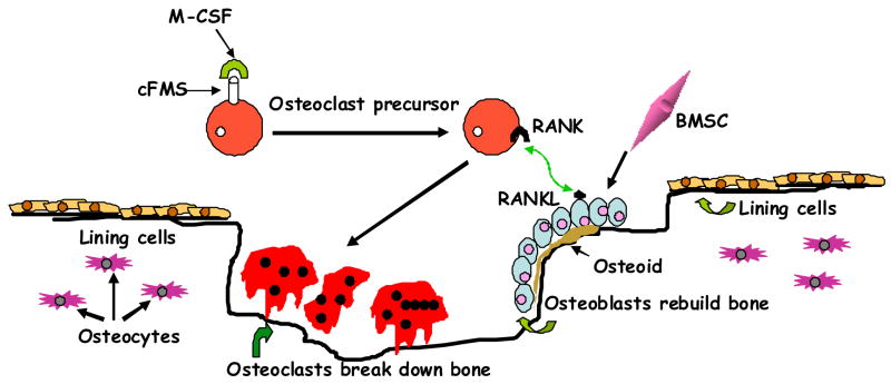
Bone remodeling. A scheme illustrating how the osteoclasts derive from hematopoietic stem cells through coordinated interaction of diverse factors; and osteoblasts from bone marrow stromal cells. The initial organic matrix (osteoid) is later calcified to form mature bone within the cavity created by osteoclasts with inclusion of osteoblasts as osteocytes within lacunae and lining cells. BMSC; bone marrow stromal cell; RANK, receptor activator of nuclear factor-κB ligand; RANKL, RANK ligand; M-CSF, macrophage-colony-stimulating factor; cFMS, cell-surface receptor in hematopoietic precursors.
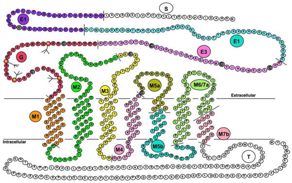
Primary sequence of the rat PTH/PTHrP receptor [9,11]. Different colors represent sequences encoded by different exons and predicted extracellular, trans-membrane spanning and cytoplasmic domains are shown. Exon nomenclature is as follow: S for exon encoding signal peptide, E1, E2 and E3 for exons encoding the extracellular extension, M1, M2, M3, M4, M5a, M5b, M6/7a and M7b for exons encoding the trans-membrane domains and portions of their connecting loops and T for the exon encoding the cytoplasmic tail. The J-domain refers to the trans-membrane domains and the connecting loops.
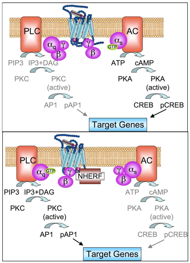
A schema for the signaling pathway regulated by NHERF. Upper panel shows the dominant cAMP signaling pathways in most cells. Lower panel illustrates how NHERF shift signaling from adenylate cyclase (AC) dominance to phospholipace C (PLC) dominance.
References
-
- Takahashi N, Udagawa N, Takami M, Suda T. Principles of Bone Biology. Academic Press; San Diego: 2002. pp. 109–126.
-
- Schluter KD. News Physiol Sci. 1999;14:243–249. - PubMed
-
- Juppner H, Abou-Samra AB, Freeman M, Kong XF, Schipani E, Richards J, Kolakowski LF, Jr, Hock J, Potts JT, Jr, Kronenberg HM, et al. Science. 1991;254(5034):1024–1026. - PubMed
-
- Karaplis AC, Luz A, Glowacki J, Bronson RT, Tybulewicz VL, Kronenberg HM, Mulligan RC. Genes Dev. 1994;8(3):277–289. - PubMed
Publication types
MeSH terms
Substances
Grants and funding
LinkOut - more resources
Full Text Sources
Research Materials
