

Widespread neocortical abnormalities in temporal lobe epilepsy with and without mesial sclerosis
- PMID: 19249372
- PMCID: PMC2799165
- DOI: 10.1016/j.neuroimage.2009.02.020
Widespread neocortical abnormalities in temporal lobe epilepsy with and without mesial sclerosis
Abstract
Purpose: Extrafocal structural abnormalities have been consistently described in temporal lobe epilepsy (TLE) with mesial temporal lobe sclerosis (TLE-MTS). In TLE without MTS (TLE-no) extrafocal abnormalities are more subtle and often require region of interest analyses for their detection. Cortical thickness measurements might be better suited to detect such subtle abnormalities than conventional whole brain volumetric techniques which are often negative in TLE-no. The aim of this study was to seek and characterize patterns of cortical thinning in TLE-MTS and TLE-no.
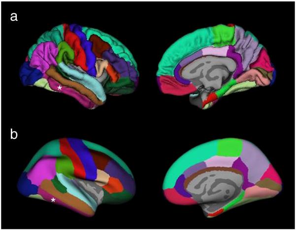
Methods: T1 weighted whole brain images were acquired on a 4 T magnet in 66 subjects (35 controls, 15 TLE-MTS, 16 TLE-no). Cortical thickness measurements were obtained using the FreeSurfer software routine. Group comparisons and correlation analyses were done using the statistical routine of FreeSurfer (FDR, p=0.05).
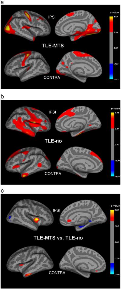
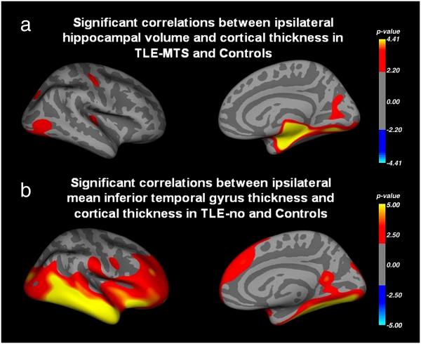
Results: TLE-MTS and TLE-no showed both widespread temporal and extratemporal cortical thinning. In TLE-MTS, the inferior medial and posterior temporal regions were most prominently affected while lateral temporal and opercular regions were more affected in TLE-no. The correlation analysis showed a significant correlation between the ipsilateral hippocampal volume and regions of thinning in TLE-MTS and between inferior temporal cortical thickness and thinning in extratemporal cortical regions in TLE-no.
Conclusion: The pattern of thinning in TLE-no was different from the pattern in TLE-MTS. This finding suggests that different epileptogenic networks could be involved in TLE-MTS and TLE and further supports the hypothesis that TLE-MTS and TLE-no might represent two distinct TLE syndromes.
Figures
References
-
- Bernasconi N, Bernasconi A, Caramanos Z, Dubeau F, Richardson J, Andermann F, et al. Entorhinal cortex atrophy in epilepsy patients exhibiting normal hippocampal volumes. Neurology. 2001;56:1335–1339. - PubMed
-
- Bernhardt BC, Worsley KJ, Besson P, Concha L, Lerch JP, Evans AC, et al. Mapping limbic network organization in temporal lobe epilepsy using morphometric correlations: Insight on the relation between mesiotemporal connectivity and cortical atrophy. NeuroImage. 2008;42(2):515–24. - PubMed
-
- Bertram EH. Why does surgery fail to cure limbic epilepsy? Seizure functional anatomy may hold the answer. Epilepsy Res. 2003;56:93–99. - PubMed
-
- Blumenfeld H, Rivera M, McNally KA, Davis K, Spencer DD, Spencer SS. Ictal neocortical slowing in temporal lobe epilepsy. Neurology. 2004;63:1015–1021. - PubMed
-
- Blumenfeld H, McNally KA, Vanderhill SD, Paige AL, Chung R, Davis K, et al. Positive and negative network correlations in temporal lobe epilepsy. Cerebral Cortex. 2004;14:892–902. - PubMed
Publication types
MeSH terms
Grants and funding
LinkOut - more resources
Full Text Sources
Medical
