. 2009 Apr 25;387(1):16-28.
doi: 10.1016/j.virol.2009.01.041. Epub 2009 Feb 27.

Mamu-A01/K(b) transgenic and MHC Class I knockout mice as a tool for HIV vaccine development

Affiliations

Mamu-A01/K(b) transgenic and MHC Class I knockout mice as a tool for HIV vaccine development

Jinliang Li et al. Virology. .

Abstract

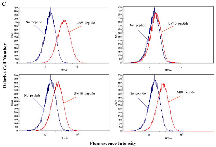
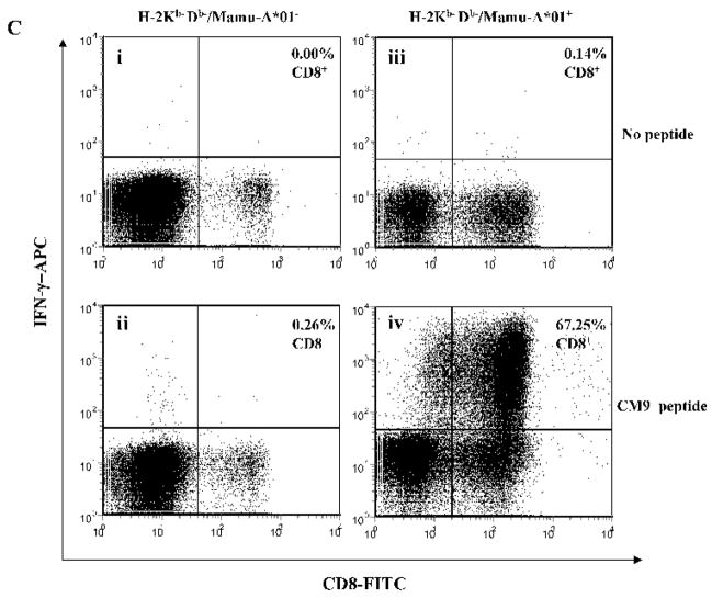
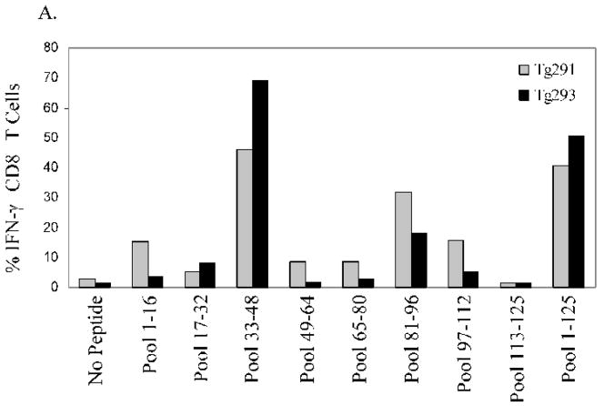
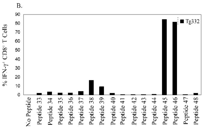
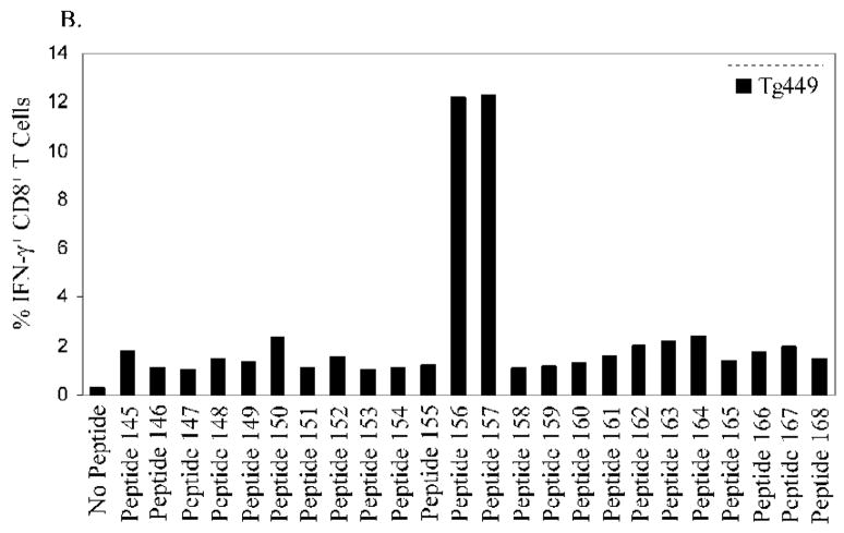
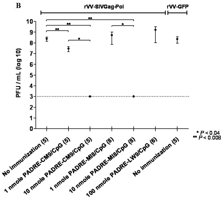
We have developed a murine model expressing the rhesus macaque (RM) Mamu-A01 MHC allele to characterize immune responses and vaccines based on antigens of importance to human disease processes. Towards that goal, transgenic (Tg) mice expressing chimeric RM (alpha1 and alpha2 Mamu-A01 domains) and murine (alpha3, transmembrane, and cytoplasmic H-2K(b) domains) MHC Class I molecules were derived by transgenesis of the H-2K(b)D(b) double MHC Class I knockout strain. After immunization of Mamu-A01/K(b) Tg mice with rVV-SIVGag-Pol, the mice generated CD8(+) T-cell IFN-gamma responses to several known Mamu-A01 restricted epitopes from the SIV Gag and Pol antigen sequence. Fusion peptides of highly recognized CTL epitopes from SIV Pol and Gag and a strong T-help epitope were shown to be immunogenic and capable of limiting an rVV-SIVGag-Pol challenge. Mamu-A01/K(b) Tg mice provide a model system to study the Mamu-A01 restricted T-cell response for various infectious diseases which are applicable to a study in RM.


Figures

Figure 1
Figure 1
(A) Scheme for generating H-2Kb promoter Mamu-A*01/Kb transgene construct. P1 and P2 are sites of Mamu-A*01 specific primers. See Materials and Methods for details of the construction of the chimeric gene. The stippled boxes correspond to exons which encode the TM and cytoplasmic (CT) domains of the H-2Kb gene (i). Clear boxes represent introns and the endogenous promoter segment is designated by a box containing an arrow indicating the direction of transcription (i). The leader peptide (LP), α1 and α2 domains of the cloned MamuA*01 MHC gene are shown as represented in the genome (ii), and their position between Nru I and Bam HI sites of the modified H-2Kb gene that was introduced by transgenesis into the DKO mice (iii). (B) Stabilization of cell surface expression of Mamu-A*01/Kb in RMA-S cells with CM9 peptide or (C) four known Mamu-A*01 restricted SIV peptides LA9, LV10, GM10 and MI8. See Table 1 for amino acid sequences. After overnight culture of Mamu-A*01/Kb-RMA-S cells at 26° C, 10−5 M of the indicated peptides were added or no peptide for the control, with subsequent incubation for 4 hrs at 37° C. Analysis was initiated by staining with MAb W6/32 conjugated to FITC, followed by flow cytometry. The GYKDGNEYI peptide represents an irrelevant peptide (control peptide).
Figure 1
Figure 1
(A) Scheme for generating H-2Kb promoter Mamu-A*01/Kb transgene construct. P1 and P2 are sites of Mamu-A*01 specific primers. See Materials and Methods for details of the construction of the chimeric gene. The stippled boxes correspond to exons which encode the TM and cytoplasmic (CT) domains of the H-2Kb gene (i). Clear boxes represent introns and the endogenous promoter segment is designated by a box containing an arrow indicating the direction of transcription (i). The leader peptide (LP), α1 and α2 domains of the cloned MamuA*01 MHC gene are shown as represented in the genome (ii), and their position between Nru I and Bam HI sites of the modified H-2Kb gene that was introduced by transgenesis into the DKO mice (iii). (B) Stabilization of cell surface expression of Mamu-A*01/Kb in RMA-S cells with CM9 peptide or (C) four known Mamu-A*01 restricted SIV peptides LA9, LV10, GM10 and MI8. See Table 1 for amino acid sequences. After overnight culture of Mamu-A*01/Kb-RMA-S cells at 26° C, 10−5 M of the indicated peptides were added or no peptide for the control, with subsequent incubation for 4 hrs at 37° C. Analysis was initiated by staining with MAb W6/32 conjugated to FITC, followed by flow cytometry. The GYKDGNEYI peptide represents an irrelevant peptide (control peptide).
Figure 2
Figure 2
(A) Detection of Mamu-A*01 DNA in tail samples from 13 Tg mice by PCR. Cont symbolizes control transgene Mamu-A*01/Kb plasmid DNA template. M represents 1 kb DNA ladder molecular size marker. (B) Detection of cell surface expression of Mamu-A*01/Kb MHC antigen in spleen cells. Splenocytes from DKO (left) mice and Mamu-A*01/Kb Tg (right) mice were stained with FITC-conjugated MAb W6/32 followed by flow cytometry. Splenocytes were also treated with 15 ng/ml IFN-γ for 16 hrs to enhance MHC antigen expression levels.
Figure 3
Figure 3
(A) Schematic map of plasmid transfer vector pSC11-GFP-SIVGag-Pol. It contains two flanking regions; TK(L) and TK(R) which are homologous to the VV-TK gene, the screen marker gene GFP under the control of the VV promoter P11, and the SIVGag-Pol gene from SIVmac239 under control of VV promoter P7.5. rVV-SIVGag-Pol was generated by transfecting pSC11-GFP-SIVGagPol into VV-infected Hu TK cells to promote homologous recombination. (B) Western Blot detection of SIV Gag protein in rVV-SIVGag-Pol infected cells. Lysates from rVV-SIVGag-Pol and wild type VV infected Hu TK cells were separated on SDS-PAGE and detected with anti-P27 MAb, followed by an HRP conjugated anti-mouse secondary antibody contained in the ECL™ kit (GE Healthcare). (C) ICC of IFN-γ production in CD8+ T lymphocytes from DKO mice (ii) and Mamu-A*01/Kb Tg mice (iv) after IVS and restimulation with CM9 peptide. Plots (i) and (iii) are negative controls without CM9 peptide restimulation. (D) Detection of IFN-γ production in CD8+ T lymphocytes after IVS with four known Mamu-A*01 specific SIV peptides: LA9 (i), GM10 (ii), LV10 (iii) and MI8 (iv). In C and D, IVS followed by overnight peptide restimulation was performed according to M&M followed by staining with anti-CD8 and anti-IFN-γ antibodies, and analyzed by flow cytometry.
Figure 3
Figure 3
(A) Schematic map of plasmid transfer vector pSC11-GFP-SIVGag-Pol. It contains two flanking regions; TK(L) and TK(R) which are homologous to the VV-TK gene, the screen marker gene GFP under the control of the VV promoter P11, and the SIVGag-Pol gene from SIVmac239 under control of VV promoter P7.5. rVV-SIVGag-Pol was generated by transfecting pSC11-GFP-SIVGagPol into VV-infected Hu TK cells to promote homologous recombination. (B) Western Blot detection of SIV Gag protein in rVV-SIVGag-Pol infected cells. Lysates from rVV-SIVGag-Pol and wild type VV infected Hu TK cells were separated on SDS-PAGE and detected with anti-P27 MAb, followed by an HRP conjugated anti-mouse secondary antibody contained in the ECL™ kit (GE Healthcare). (C) ICC of IFN-γ production in CD8+ T lymphocytes from DKO mice (ii) and Mamu-A*01/Kb Tg mice (iv) after IVS and restimulation with CM9 peptide. Plots (i) and (iii) are negative controls without CM9 peptide restimulation. (D) Detection of IFN-γ production in CD8+ T lymphocytes after IVS with four known Mamu-A*01 specific SIV peptides: LA9 (i), GM10 (ii), LV10 (iii) and MI8 (iv). In C and D, IVS followed by overnight peptide restimulation was performed according to M&M followed by staining with anti-CD8 and anti-IFN-γ antibodies, and analyzed by flow cytometry.
Figure 4
Figure 4
Mapping Mamu-A*01 restricted epitopes of SIV Gag by intracellular IFN-γ staining in CD8+ T cells after IVS. Splenocytes from rVV-SIVGag-Pol immunized Mamu-A*01/Kb Tg mice were subjected to IVS with 8 SIV Gag peptide subpools or the full-length peptide library. (A) Reactive peptide pools were deconvoluted by testing individual SIV Gag peptides 33 to 48 (B), 81 to 96 (C).
Figure 4
Figure 4
Mapping Mamu-A*01 restricted epitopes of SIV Gag by intracellular IFN-γ staining in CD8+ T cells after IVS. Splenocytes from rVV-SIVGag-Pol immunized Mamu-A*01/Kb Tg mice were subjected to IVS with 8 SIV Gag peptide subpools or the full-length peptide library. (A) Reactive peptide pools were deconvoluted by testing individual SIV Gag peptides 33 to 48 (B), 81 to 96 (C).
Figure 5
Figure 5
Mapping Mamu-A*01 restricted epitopes of SIV Pol by intracellular IFN-γ staining in CD8+ T cells after IVS. Splenocytes from rVV-SIVGag-Pol immunized Mamu-A*01/Kb Tg mice were subjected to IVS in similar conditions as described in the legend to Figure 4 with 11 SIV Pol peptide subpools (A). Individual SIV Pol peptide pools were evaluated between 145 to 168 (B).
Figure 5
Figure 5
Mapping Mamu-A*01 restricted epitopes of SIV Pol by intracellular IFN-γ staining in CD8+ T cells after IVS. Splenocytes from rVV-SIVGag-Pol immunized Mamu-A*01/Kb Tg mice were subjected to IVS in similar conditions as described in the legend to Figure 4 with 11 SIV Pol peptide subpools (A). Individual SIV Pol peptide pools were evaluated between 145 to 168 (B).
Figure 6
Figure 6
Immunogenicity (A) and challenge studies (B) conducted with fusion peptide immunized Tg mice. (A) Tg mice (N=2) were immunized with 100 nmoles of fusion peptides + 25 μg CPG 7909 DNA one time, and were sacrificed after 14 days for processing of splenocytes for ICC studies. ICC assays were carried out as described in M&M using CTL epitope encoded by fusion peptide (filled bars) or without exogenous peptide (unfilled bars). (B) Tg mice (number of mice indicated in parentheses for each group on the x-axis) were immunized with indicated amounts of fusion peptides + 25 μg CPG 7909 DNA twice as described in M&M. Seven days later, rVV-SIVGag-Pol or rVV-GFP were administered i.p. to immunized mice, as indicated in the legend above each data series. Five days later, mice were sacrificed, ovary lysates prepared, and diluted before incubating with monolayers of CV-1 cells as described in M&M. Plaques were visualized using immunostain to VV antigens as described in M&M. Dotted line indicates the limit of detection of VV plaques using the assay. P-values (2-sided) were determined using the Wilcoxon rank-sum test, and values which are significant are shown as one (P<0.04) or two asterisks (P<0.008). Standard error of the mean is shown as horizontal bars, with means indicated as filled circles. Undetectable plaques in all members of a group are represented by a single filled circle without error bars on the dotted line signifying the minimal detection limit.
Figure 6
Figure 6
Immunogenicity (A) and challenge studies (B) conducted with fusion peptide immunized Tg mice. (A) Tg mice (N=2) were immunized with 100 nmoles of fusion peptides + 25 μg CPG 7909 DNA one time, and were sacrificed after 14 days for processing of splenocytes for ICC studies. ICC assays were carried out as described in M&M using CTL epitope encoded by fusion peptide (filled bars) or without exogenous peptide (unfilled bars). (B) Tg mice (number of mice indicated in parentheses for each group on the x-axis) were immunized with indicated amounts of fusion peptides + 25 μg CPG 7909 DNA twice as described in M&M. Seven days later, rVV-SIVGag-Pol or rVV-GFP were administered i.p. to immunized mice, as indicated in the legend above each data series. Five days later, mice were sacrificed, ovary lysates prepared, and diluted before incubating with monolayers of CV-1 cells as described in M&M. Plaques were visualized using immunostain to VV antigens as described in M&M. Dotted line indicates the limit of detection of VV plaques using the assay. P-values (2-sided) were determined using the Wilcoxon rank-sum test, and values which are significant are shown as one (P<0.04) or two asterisks (P<0.008). Standard error of the mean is shown as horizontal bars, with means indicated as filled circles. Undetectable plaques in all members of a group are represented by a single filled circle without error bars on the dotted line signifying the minimal detection limit.

Similar articles

Cited by

References

    1. Ahlers JD, Belyakov IM, Matsui S, Berzofsky JA. Mechanisms of cytokine synergy essential for vaccine protection against viral challenge. Int Immunol. 2001;13:897–908. - PubMed
    1. Alexander J, Sidney J, Southwood S, Ruppert J, Oseroff C, Maewal A, Snoke K, Serra HM, Kubo RT, Sette A. Development of high potency universal DR-restricted helper epitopes by modification of high affinity DR-blocking peptides. Immunity. 1994;1:751–761. - PubMed
    1. Allen TM, Mothe BR, Sidney J, Jing P, Dzuris JL, Liebl ME, Vogel TU, O’Connor DH, Wang X, Wussow MC, Thomson JA, Altman JD, Watkins DI, Sette A. CD8(+) lymphocytes from simian immunodeficiency virus-infected rhesus macaques recognize 14 different epitopes bound by the major histocompatibility complex class I molecule mamu-A*01: implications for vaccine design and testing. The Journal of Virology. 2001;75:738–749. - PMC - PubMed
    1. Allen TM, Sidney J, Del Guercio MF, Glickman RL, Lensmeyer GL, Wiebe DA, DeMars R, Pauza CD, Johnson RP, Sette A, Watkins DI. Characterization of the peptide binding motif of a rhesus MHC class I molecule (Mamu-A*01) that binds an immunodominant CTL epitope from simian immunodeficiency virus. J Immunol. 1998;160:6062–6071. - PubMed
    1. Arnold B, Hammerling GJ. MHC class-I transgenic mice. Annu Rev Immunol. 1991;9:297–322. 297–322. - PubMed

Publication types

MeSH terms