. 2009 Feb 27;33(4):483-95.
doi: 10.1016/j.molcel.2009.01.011.

E2-RING expansion of the NEDD8 cascade confers specificity to cullin modification

Affiliations

E2-RING expansion of the NEDD8 cascade confers specificity to cullin modification

Danny T Huang et al. Mol Cell. .

Abstract

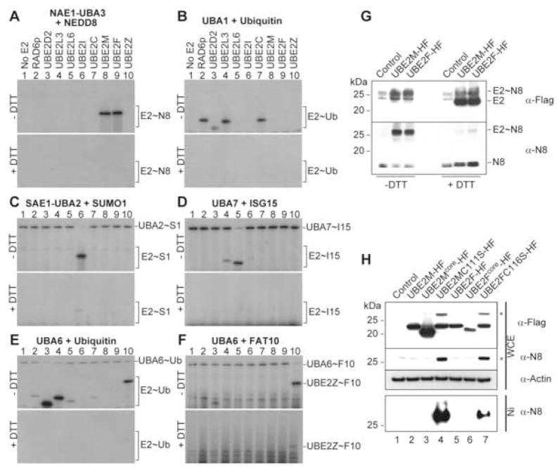
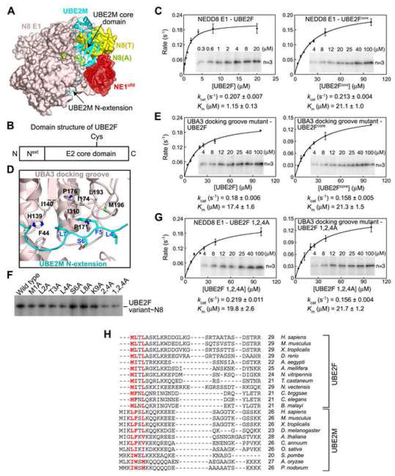
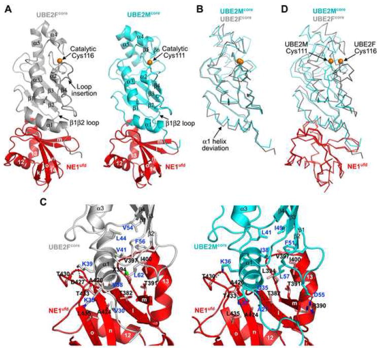
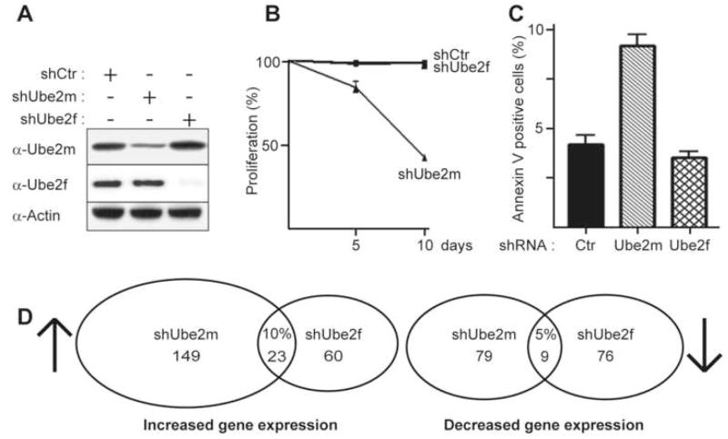
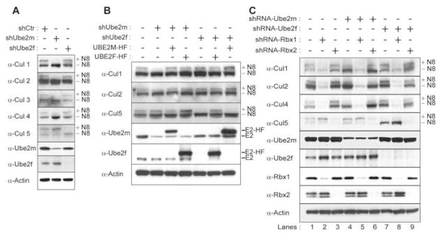
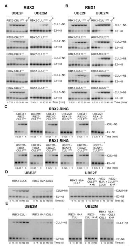
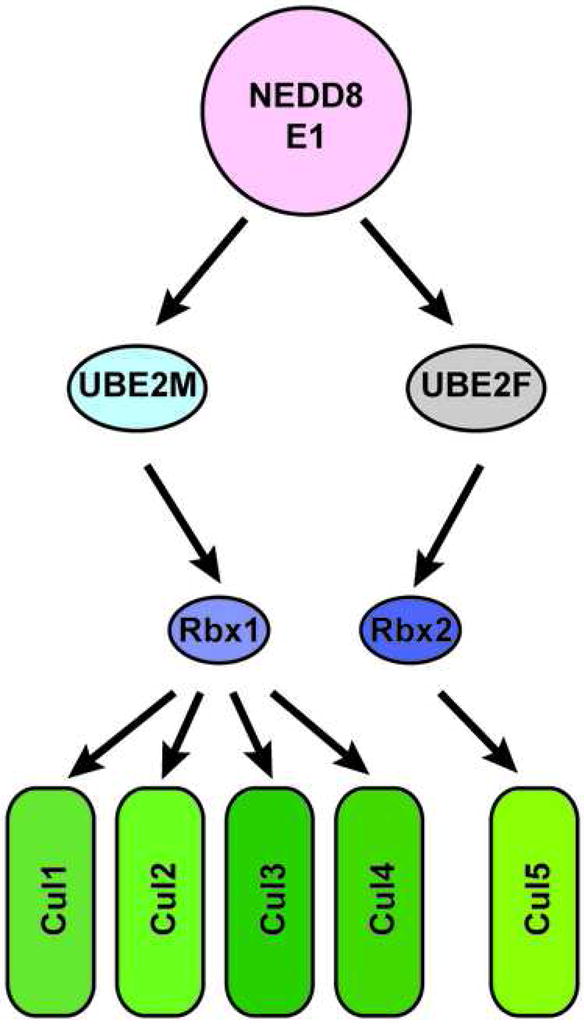
Ubiquitin and ubiquitin-like proteins (UBLs) are directed to targets by cascades of E1, E2, and E3 enzymes. The largest ubiquitin E3 subclass consists of cullin-RING ligases (CRLs), which contain one each of several cullins (CUL1, -2, -3, -4, or -5) and RING proteins (RBX1 or -2). CRLs are activated by ligation of the UBL NEDD8 to a conserved cullin lysine. How is cullin NEDD8ylation specificity established? Here we report that, like UBE2M (also known as UBC12), the previously uncharacterized E2 UBE2F is a NEDD8-conjugating enzyme in vitro and in vivo. Biochemical and structural analyses indicate how plasticity of hydrophobic E1-E2 interactions and E1 conformational flexibility allow one E1 to charge multiple E2s. The E2s have distinct functions, with UBE2M/RBX1 and UBE2F/RBX2 displaying different target cullin specificities. Together, these studies reveal the molecular basis for and functional importance of hierarchical expansion of the NEDD8 conjugation system in establishing selective CRL activation.


Figures

Figure 1
Figure 1. UBE2F forms a NEDD8 thioester conjugate in vitro and in vivo.
Autoradiograms showing 32P-UBL thioesters after E2 charging by E1~32P-UBLs (top) and reduction with 100 mM DTT (bottom): A, NAE1-UBA3~NEDD8; B, UBA1~Ub; C, SAE1-UBA2~SUMO1; D, UBA7~ISG15; E, UBA6~Ub; F, UBA6~ FAT10. G, Immunoblot showing 100 mM DTT reduction of E2~NEDD8 complex detected with antibodies against E2 (α-Flag) and NEDD8 (α-N8) from NIH 3T3 cells infected with indicated retroviruses. H, Immunoblot as in G (top) from whole cell extracts (WCE) of NIH 3T3 cells infected with indicated retroviruses and lysed in urea buffer, and from nickel-agarose pull-downs (His). * expected MW of E2~NEDD8 oxy-ester complex.
Figure 2
Figure 2. Bipartite UBE2F interaction with UBA3: a conserved Φ-Φ-X-Φ UBA3 in NEDD8 E2 N-extensions
A, Structure of NAE1 UBA3~NEDD8(T)-NEDD8(A)-MgATP-UBE2M(C111A) (Huang et al., 2007) depicting interactions between NEDD8’s E1 (NAE1-UBA3, light pink, with ufd in red) and UBE2M (cyan). The thioester-bound NEDD8 (N8(T)) is yellow and adenylation site NEDD8 (N8(A)) is lime. UBE2M’s N-extension and core domains are indicated. B, Domain structure of UBE2F. C, Michaelis-Menten curves for 32P-NEDD8 thioester conjugate formation by UBE2F and UBE2Fcore. D, Detailed interactions between UBE2M’s N-extension (blue) and UBA3’s docking groove (black) (Huang et al., 2004). E, Michaelis-Menten curves for UBA3 docking groove mutant-catalyzed 32P-NEDD8 thioester conjugate formation by UBE2F and UBE2Fcore. F, Autoradiogram of 32P-NEDD8 thioester conjugates formed by the indicated Ala mutant of UBE2F. G, Michaelis-Menten curves for 32P-NEDD8 thioester conjugate formation by UBE2F and NAE1-UBA3 and mutants. H, Structure-based sequence alignment of UBE2F and UBE2M’s N-terminal extension from various species (Tables S2, S3). Kinetic analyses (C, E, G) show standard errors, insets are representative autoradiograms indicating E2 concentrations and the number of independent replicates, and kinetic constants (kcat and Km values) are indicated.
Figure 3
Figure 3. Structural basis for NEDD8 E1 ufd’s binding to varying E2 sequences
A, Overall structure of NE1ufd-UBE2Fcore (left) compared to NE1ufd-UBE2Mcore (Huang et al., 2005) (right). NE1ufd is red, UBE2Fcore grey, UBE2Mcore cyan, and E2 catalytic cysteines orange spheres. B, UBE2Fcore and UBE2Mcore structural superposition. C, Close-ups of NE1ufd-UBE2Fcore (left) and NE1ufd-UBE2Mcore (right) interfaces. Oxygens are red, nitrogens blue, ionic interactions dashed. E2 residues are labeled in blue, and NE1ufd’s in black. X – Leu394SeMet. D, Structural superposition of the NE1ufd portions of NE1ufd-UBE2Fcore and NE1ufd-UBE2Mcore.
Figure 4
Figure 4. Effects of Ube2f and Ube2m knockdown on global cellular properties
A, Immunoblots of Ube2f and Ube2m proteins after infecting NIH 3T3 cells with retroviruses expressing shRNAs for control (Ctr), Ube2m or Ube2f. B, Average % of cells in culture (3 independent experiments) relative to control 5 and 10 days after infecting NIH 3T3 cells with the indicated retroviruses. C, Average % of Annexin V positive cells (3 independent experiments) detected by flow cytometry 8 days after infecting NIH 3T3 cells with the indicated retroviruses. Error bars – standard error. D, Venn diagram displaying the results of transcriptome analysis from 3 independent experiments. Total numbers of up- and down-regulated genes for NIH 3T3 cells treated with shRNA against Ube2m or Ube2f versus control, displaying % overlap.
Figure 5
Figure 5. Distinct E2-Rbx pairs regulate cullin NEDD8ylation specificity in vivo-N8 indicates unNedd8ylated and +N8 indicates Nedd8ylated cullin
A, Immunoblots of urea lysates from NIH 3T3 cells infected with retroviruses expressing shRNA against control (Ctr), Ube2m or Ube2f, probed with the indicated antisera. B, Immunoblots of urea lysates from retrovirally infected NIH 3T3 cells as in A, in the absence or presence of retroviruses expressing human UBE2M-HF or human UBE2F-HF, probed with the indicated antisera. C, Immunoblots of urea lysates from retrovirally infected NIH 3T3 cells as in A, in the absence or presence of retroviruses expressing shRNA against Rbx1 or Rbx2, probed with the indicated antisera.
Figure 6
Figure 6. Combinatorial roles of RBXs and E2s establish cullin NEDD8ylation specificity in vitro
Autoradiograms of chase reactions monitoring A, RBX1- or B, RBX2-mediated 32P-NEDD8 transfer from UBE2F (left) and UBE2M (right) to the indicated CULCTD. C, Autoradiograms for chase reactions monitoring 32P-NEDD8 transfer from UBE2F or UBE2M to the indicated CULCTD mediated by RBX chimeras harboring the indicated N-terminus/RBX2 RING (top panel), and the indicated N-terminus/RBX1 RING (bottom panel). D, UBE2F reactions as in A and B, except with RBX2(Ile52Ala)-CUL5 (left) and wild-type RBX2 in complex with non-NEDD8ylatable CUL5CTD(K724R) as indicated (right). E, UBE2M reactions as in A and B, except with RBX2(Ile44Ala)-CUL1 (left) and wild-type RBX1 in complex with non-NEDD8ylatable CUL1CTD(K720R) as indicated (right).
Figure 7
Figure 7. Hierarchical E2-RING expansion of the NEDD8 cascade
Schematic view of distinct molecular pathways dictating specificity of cullin NEDD8ylation.

References

    1. The CCP4 suite: programs for protein crystallography. Acta crystallographica. 1994;50:760–763. - PubMed
    1. Amir RE, Iwai K, Ciechanover A. The NEDD8 pathway is essential for SCF(beta -TrCP)-mediated ubiquitination and processing of the NF-kappa B precursor p105. The Journal of biological chemistry. 2002;277:23253–23259. - PubMed
    1. Brzovic PS, Keeffe JR, Nishikawa H, Miyamoto K, Fox D, 3rd, Fukuda M, Ohta T, Klevit R. Binding and recognition in the assembly of an active BRCA1/BARD1 ubiquitin-ligase complex. Proceedings of the National Academy of Sciences of the United States of America. 2003;100:5646–5651. - PMC - PubMed
    1. Cardozo T, Pagano M. The SCF ubiquitin ligase: insights into a molecular machine. Nature reviews. 2004;5:739–751. - PubMed
    1. Chiu YH, Sun Q, Chen ZJ. E1-L2 activates both ubiquitin and FAT10. Molecular cell. 2007;27:1014–1023. - PubMed

Publication types

Associated data