. 2009 Feb 27;323(5918):1208-1211.
doi: 10.1126/science.1165942.

Mutations in FUS, an RNA processing protein, cause familial amyotrophic lateral sclerosis type 6

Affiliations

Mutations in FUS, an RNA processing protein, cause familial amyotrophic lateral sclerosis type 6

Caroline Vance et al. Science. .

Abstract

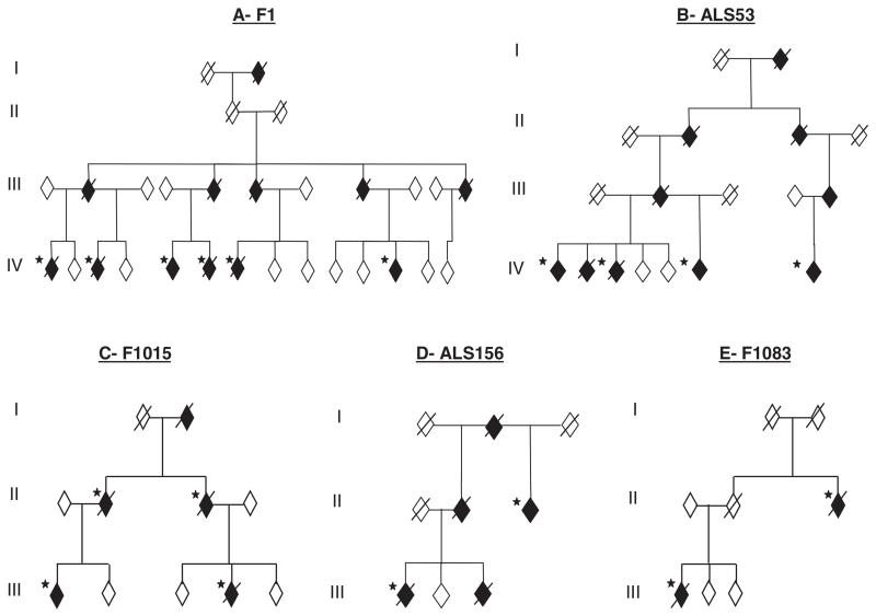
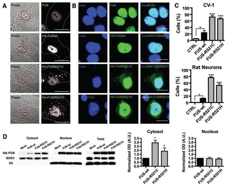
Amyotrophic lateral sclerosis (ALS) is a fatal neurodegenerative disease that is familial in 10% of cases. We have identified a missense mutation in the gene encoding fused in sarcoma (FUS) in a British kindred, linked to ALS6. In a survey of 197 familial ALS index cases, we identified two further missense mutations in eight families. Postmortem analysis of three cases with FUS mutations showed FUS-immunoreactive cytoplasmic inclusions and predominantly lower motor neuron degeneration. Cellular expression studies revealed aberrant localization of mutant FUS protein. FUS is involved in the regulation of transcription and RNA splicing and transport, and it has functional homology to another ALS gene, TARDBP, which suggests that a common mechanism may underlie motor neuron degeneration.


Figures

Fig. 1
Fig. 1
FUS mutations segregate with disease in five dominant ALS kindreds. Affected individuals are indicated by black symbols and unaffected individuals by open symbols. Slashed symbols indicate deceased individuals. Asterisks indicates an affected individual for whom DNA was available. All affected individuals tested carried their families’ mutation. Gender, birth order, and the mutation status of unaffected individuals have been omitted for reasons of confidentiality.
Fig. 2
Fig. 2
Patients with FUS mutations develop cytoplasmic FUS immunoreactive inclusions in lower motor neurons. Antibody to FUS immunolabels inclusions within the anterior horn (dotted line) of the spinal cord in patients with FUS mutations (A to F and arrowheads in O). Staining in controls (G, H, and M), mutant SOD1 FALS (I and J), and SALS (K, L, and N) demonstrate diffuse nuclear staining (arrows) with variable intensity without inclusions. Scale bars, 12 μm [(A) to (L)], 50 μm [(M) to (O)].
Fig. 3
Fig. 3
FUS mutations cause subcellular mislocalization. (A and B) The subcellular localization of endogenous FUS, transfected wild-type (HA-FUSWT), and FUS mutants (HA-FUSR521C, HA-FUSR521H) was determined in CV-1 cells (A) and rat cortical neurons (B) by immunofluorescence. The plasma membrane (A, yellow dash) and nucleus (A, red dash) of representative CV-1 cells are outlined on phase contrast (A, left panel) and corresponding FUS immunofluorescence images (A, right panel). Nuclear diamidino-2-phenylindole (DAPI) staining (B, left panel), FUS immunofluorescence (B, middle panel), and overlay (B, right panel) of representative cortical neurons are shown. Scale bars, 25 μm (A), 10 μm (B). (C) The percentage of CV-1 cells (upper panel) and cortical neurons (lower panel) showing FUS staining in the cytoplasm was significantly increased for mutant forms of FUS compared to wild-type and endogenous. Statistical significance was determined by one-way analysis of variance (P < 0.0001 for CV1 and neurons) followed by Bonferroni’s Multiple Comparison Test (*, P < 0.05; ***, P <0.001). (D) FUS mutants show a significant increase in the cytosolic fraction. N2A cells transfected with FUSWT, FUSR521C, and FUSR521H were separated into cytosolic and nuclear fractions, and the amount of transfected FUS was determined by densitometry of antibody to HA immunoblots (mean ± SEM, n = 3). The purity of fractions was determined with antibody to SOD1 (cytosol) and antibody to histone H3 (nucleus), and the transfection efficiencies were compared in total cell lysates. Statistical significance was determined by Wilcoxon Signed Rank Test (*, P < 0.05).

References

    1. Shaw CE, al-Chalabi A, Leigh N. Curr. Neurol. Neurosci. Rep. 2001;1:69. - PubMed
    1. Shaw CE, et al. Neurology. 1997;49:1612. - PubMed
    1. Rosen DR. Nature. 1993;364:362. - PubMed
    1. Conforti FL, et al. Neuromuscul. Disord. 2008;18:68. - PubMed
    1. Greenway MJ, et al. Nat. Genet. 2006;38:411. - PubMed

Publication types

MeSH terms

Substances