. 2009 Feb 27;323(5918):1211-5.
doi: 10.1126/science.1169096.

Synchronous hyperactivity and intercellular calcium waves in astrocytes in Alzheimer mice

Affiliations

Synchronous hyperactivity and intercellular calcium waves in astrocytes in Alzheimer mice

Kishore V Kuchibhotla et al. Science. .

Abstract

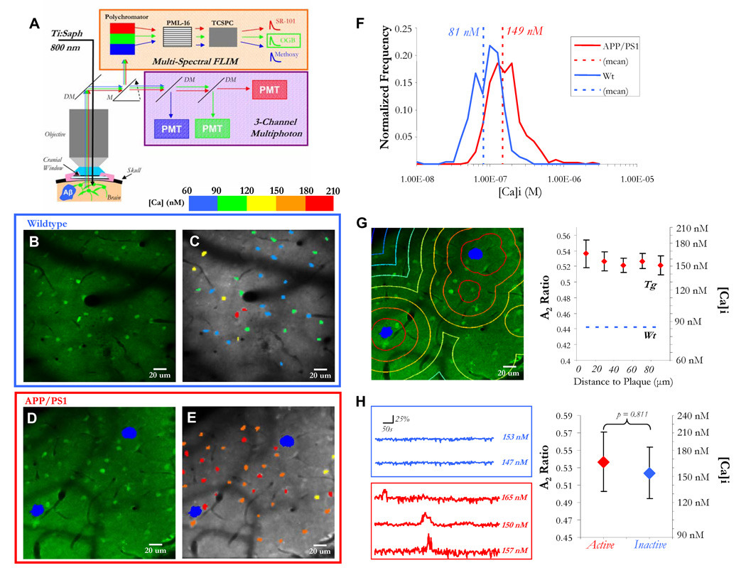
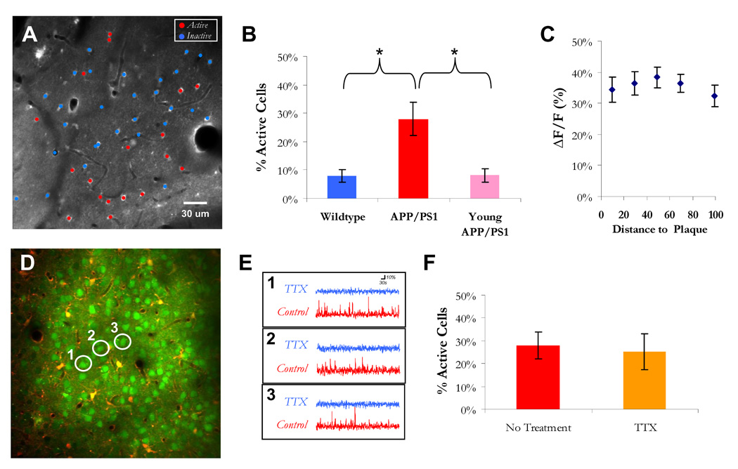
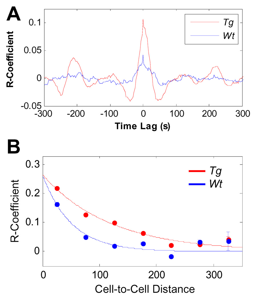
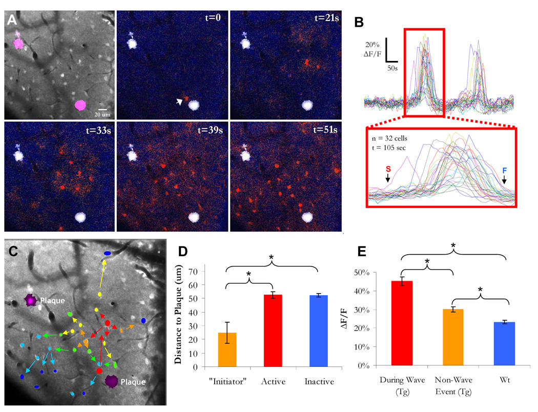
Although senile plaques focally disrupt neuronal health, the functional response of astrocytes to Alzheimer's disease pathology is unknown. Using multiphoton fluorescence lifetime imaging microscopy in vivo, we quantitatively imaged astrocytic calcium homeostasis in a mouse model of Alzheimer's disease. Resting calcium was globally elevated in the astrocytic network, but was independent of proximity to individual plaques. Time-lapse imaging revealed that calcium transients in astrocytes were more frequent, synchronously coordinated across long distances, and uncoupled from neuronal activity. Furthermore, rare intercellular calcium waves were observed, but only in mice with amyloid-beta plaques, originating near plaques and spreading radially at least 200 micrometers. Thus, although neurotoxicity is observed near amyloid-beta deposits, there exists a more general astrocyte-based network response to focal pathology.


Figures

Figure 1
Figure 1. Resting calcium is globally elevated in astrocytic networks
A) Multiphoton laser illumination simultaneously excited methoxy-XO4 (blue, amyloid-β), OGB (green, neurons and astrocytes), and SR-101 (red, astrocytes) through a cranial window. The resulting fluorescence emission was sent to either (1) a three-channel intensity-based PMT module or (2) a 16-channel multi-spectral FLIM detector. A single-photon counter (tcSPC) recorded fluorescence lifetime data . B–E) Fluorescence decay curves were fit with a calcium bound lifetime (2359 ps) and an unbound calcium lifetime (569 ps) for each pixel. The pixel data were averaged to obtain single- cell calcium levels, depicted with a calibrated colorbar (C,E). Astrocytes in APP/PS1 mice with cortical plaques (in blue, D–E), exhibited significantly higher levels of [Ca]i than in wildtype mice (B–C,F: p < 0.05, Student’s t-test, n = 241 cells in 3 mice (wt), n = 364 cells in 3 mice (Tg)). G) Astrocyte resting [Ca]i did not depend on proximity to a plaque (p = 0.9194, Kruskal-Wallis test, n > 25 cells for each distance group). H) There was no difference in resting calcium between cells that were active versus inactive (p = 0.811, Student’s t-test, n = 209 cells in 3 mice).
Figure 2
Figure 2. Neuron-independent increase in spontaneous activity throughout astrocytic network
A) Cell activity map overlaid on multiphoton image of astrocytes. B) There were more spontaneously active cells in mice with cortical plaques (*, p < 0.05, Kruskal-Wallis with Tukey-Cramer post-hoc, n = 15 mice, 8 APP/PS1 with plaques, 4 non-transgenic and 3 APP/PS1 before plaque deposition). C) The amplitude of the [Ca]i transients (n = 160 astrocytes) did not depend on proximity to plaque (5 groups, p = 0.9178, Kruskal-Wallis test). D) Multiphoton image of neurons and astrocytes in an APP/PS1 transgenic mouse with cortical plaques. Three neurons are highlighted to show their spontaneous activity traces before and after application of 1 uM TTX in (E). F) There was no reduction in the percentage of active astrocytes in the presence of TTX (27.9 +/− 6.0% in control (n=8 mice, 1,241 astrocytes) vs. 25.4 +/− 7.8% under TTX (n = 4 mice, 818 astrocytes), p = 0.8081, Mann-Whitney U-test).
Figure 3
Figure 3. Spatiotemporal synchrony of astrocytic calcium signaling in APP/PS1 mice
A) The mean cross-correlogram for all cell pairs (excluding autocorrelations) in transgenic and non- transgenic mice. In APP/PS1 mice there was an increase in the probability that two cells had coordinated activity (n = 3 mice for APP/PS1 and Wt, n = 1,257 cell pairs in transgenic and n = 471 cell pairs in Wt). B) Cell-pair distance (x-axis) versus Correlation coefficient (y-axis). Data were fit to a mono-exponential. In transgenic mice (red), cell-pairs exhibited significantly correlated activity at distances up to 200 µm (p < 0.01, Kruskal-Wallis test with Tukey-Cramer post-hoc) whereas in wildtype mice (blue), cell-pairs were not correlated at inter-cellular distances greater than 50 µm (p = 0.65, Kruskal-Wallis with Tukey-Cramer post-hoc).
Figure 4
Figure 4. Intercellular calcium waves in mice with cortical plaques
A,B) A time-lapse ΔF/F image filmstrip and data of calcium activity in an APP/PS1 transgenic mouse. The white arrow in the second panel points to the “initiator” astrocyte and the sequential panels show the propagation of the wave. C) A summary of (A) in which color denotes temporal sequence and arrows denote spatial propagation. D) Astrocytes that “initiate” ICWs were closer to senile plaques than the average active or inactive astrocyte (*, p < 0.05, Kruskal-Wallis with Tukey-Cramer post-hoc). E) There was a significant increase in the amplitude of the calcium signal during a wave (* = p < 0.001, ANOVA with Tukey- Cramer post-hoc, n = 47 cells in Tg (wave), n = 156 cells in Tg (non-wave), n = 168 in Wt).

References

    1. Meyer-Luehmann M, et al. Nature. 2008 Feb 7;451:720. - PMC - PubMed
    1. Busche MA, et al. Science. 2008 Sep 19;321:1686. - PubMed
    1. Spires TL, et al. J Neurosci. 2005 Aug 3;25:7278. - PMC - PubMed
    1. Kuchibhotla KV, et al. Neuron. 2008 Jul 31;59:214. - PMC - PubMed
    1. Wang X, et al. Nat Neurosci. 2006 Jun;9:816. - PubMed

Publication types