. 2009 Mar 17;106(11):4319-24.
doi: 10.1073/pnas.0810343106. Epub 2009 Feb 27.

Transcription factor function and promoter architecture govern the evolution of bacterial regulons

Affiliations

Transcription factor function and promoter architecture govern the evolution of bacterial regulons

J Christian Perez et al. Proc Natl Acad Sci U S A. .

Abstract

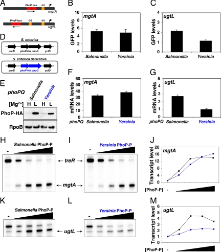
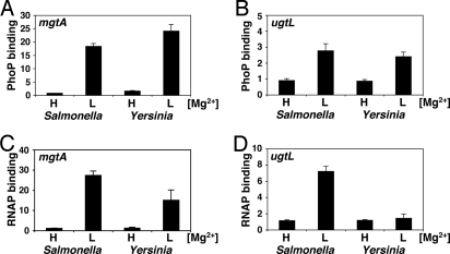
Evolutionary changes in ancestral regulatory circuits can bring about phenotypic differences between related organisms. Studies of regulatory circuits in eukaryotes suggest that these modifications result primarily from changes in cis-regulatory elements (as opposed to alterations in the transcription factors that act upon these sequences). It is presently unclear how the evolution of gene regulatory circuits has proceeded in bacteria, given the rampant effects of horizontal gene transfer, which has significantly altered the composition of bacterial regulons. We now demonstrate that the evolution of the regulons governed by the regulatory protein PhoP in the related human pathogens Salmonella enterica and Yersinia pestis has entailed functional changes in the PhoP protein as well as in the architecture of PhoP-dependent promoters. These changes have resulted in orthologous PhoP proteins that differ both in their ability to promote transcription and in their role as virulence regulators. We posit that these changes allow bacterial transcription factors to incorporate newly acquired genes into ancestral regulatory circuits and yet retain control of the core members of a regulon.


Conflict of interest statement

The authors declare no conflict of interest.

Figures

Fig. 1.
Fig. 1.
The Salmonella and Yersinia PhoP proteins differ in their ability to promote gene transcription. (A) The Salmonella mgtA and ugtL promoters differ both in the orientation and the distance of the PhoP box to the promoter −10 region. The PhoP box is located in the place that corresponds to the promoter −35 region in the mgtA promoter but is 20 bp further upstream in the ugtL promoter. (B and C) GFP expression driven by the Salmonella mgtA and ugtL promoters in Salmonella and Yersinia. Approximately 150-nt DNA fragments corresponding to the mgtA and ugtL promoter regions (covering ≈130 nucleotides upstream and ≈20 nucleotides downstream of the transcription start sites) were cloned in front of a promoterless gfp gene in the low-copy plasmid pMS201. Organisms were grown in defined medium containing 50 μM MgSO4, inducing conditions for the PhoP/PhoQ system. GFP expression was normalized to cell density. Shown are the ratios of the normalized GFP values between wild-type Salmonella (14028s) or Yersinia (KIM6) and their respective isogenic phoP mutant strains (EG15598 and EG14737, respectively). Values shown are mean plus SD of at least 3 independent experiments. (D) Schematic of the genomic context of the phoPQ locus in Salmonella strain EG13918 coding for the Salmonella PhoP-HA and PhoQ proteins and its derivative harboring the Yersinia phoP-HA and phoQ genes (in blue) (EG17569). (E) Expression of the Salmonella and Yersinia PhoP proteins in the Salmonella strains EG13918 and EG17569 depicted in D. Western blot analysis was performed with anti-HA and anti-RpoB antibodies (to detect the PhoP-HA and RpoB proteins, respectively) on cell extracts prepared from bacteria grown as described in B and C in medium containing 10 mM (H) or 50 μM (L) MgSO4. (F and G) mgtA and ugtL expression in the Salmonella strains depicted in D. Cells were grown as described in B and C. Transcript levels were determined by quantitative real-time PCR and normalized to ribosomal RNA levels. Shown are the ratios of the normalized transcript levels present in the strains described in D relative to those produced by the phoPQ mutant EG15598. Values shown are mean plus SD of at least 3 independent experiments. (H–M) Single-round in vitro transcription assays with linear templates corresponding to the mgtA (H–J) and ugtL (K–M) promoters, E. coli RNA polymerase, and increasing amounts of phosphorylated Salmonella or Yersinia PhoP proteins. The upper band in H–I corresponds to treR, a PhoP-repressed transcript going in the reverse orientation (22). The upper band in K and L corresponds to a spurious transcript observed in vitro but not in vivo (21, 45). Quantification of the in vitro transcription assays is shown in J for mgtA and in M for ugtL.
Fig. 2.
Fig. 2.
The Yersinia PhoP protein binds to the ugtL promoter but cannot recruit RNA polymerase. (A–D) Promoter occupancy by PhoP (A and B) and RNA polymerase (C and D) determined by ChIP in isogenic Salmonella strains expressing the Salmonella (EG13918) or the Yersinia (EG17569) phoPQ operon. Cells were grown in N-minimal medium containing 10 mM (H) or 10 μM (L) MgCl2. Shown are the mean plus SD of at least 3 independent experiments.
Fig. 3.
Fig. 3.
Architectures of PhoP-activated promoters in Salmonella and Yersinia. (A and B) Sequences of the promoter regions of the transcripts directly activated by PhoP in Salmonella (A) and Yersinia (B). The transcription start sites are indicated in blue, the PhoP boxes are in red, and the putative −10 sequences are underlined. Boxes and arrows indicate the location and orientation of the PhoP binding sites across promoters. The Salmonella PhoP-activated transcripts have been described previously (23, 26). The Yersinia PhoP-dependent transcripts were uncovered as described in Materials and Methods (Fig. S4). PhoP binding to all promoters was determined in vivo by ChIP-chip and/or ChIP-real-time PCR and in several cases in vitro by DNase I footprinting (Fig. S4). (C) Summary of the distribution of promoter architectures and the differential ability of the Salmonella and Yersinia PhoP proteins to activate transcription. Although the mgtA-like promoter architecture is shared by several Salmonella and Yersinia PhoP-activated genes, other members of the regulon harbor promoter structures that are absent from the other organism. The Salmonella and Yersinia PhoP proteins differ in their ability to activate expression from genes of the latter group.

References

    1. Tuch BB, Galgoczy DJ, Hernday AD, Li H, Johnson AD. The evolution of combinatorial gene regulation in fungi. PLoS Biol. 2008;6:e38. - PMC - PubMed
    1. Tuch BB, Li H, Johnson AD. Evolution of eukaryotic transcription circuits. Science. 2008;319:1797–1799. - PubMed
    1. Carroll SB. Evolution at two levels: On genes and form. PLoS Biol. 2005;3:e245. - PMC - PubMed
    1. Stern DL. Evolutionary developmental biology and the problem of variation. Evolutio (Lawrence, Kans) 2000;54:1079–1091. - PubMed
    1. Wray GA. The evolutionary significance of cis-regulatory mutations. Nat Rev Genet. 2007;8:206–216. - PubMed

Publication types

LinkOut - more resources