. 2009 Mar 17;106(11):4154-9.
doi: 10.1073/pnas.0813360106. Epub 2009 Feb 27.

Type VI secretion apparatus and phage tail-associated protein complexes share a common evolutionary origin

Affiliations

Type VI secretion apparatus and phage tail-associated protein complexes share a common evolutionary origin

Petr G Leiman et al. Proc Natl Acad Sci U S A. .

Abstract

Protein secretion is a common property of pathogenic microbes. Gram-negative bacterial pathogens use at least 6 distinct extracellular protein secretion systems to export proteins through their multilayered cell envelope and in some cases into host cells. Among the most widespread is the newly recognized Type VI secretion system (T6SS) which is composed of 15-20 proteins whose biochemical functions are not well understood. Using crystallographic, biochemical, and bioinformatic analyses, we identified 3 T6SS components, which are homologous to bacteriophage tail proteins. These include the tail tube protein; the membrane-penetrating needle, situated at the distal end of the tube; and another protein associated with the needle and tube. We propose that T6SS is a multicomponent structure whose extracellular part resembles both structurally and functionally a bacteriophage tail, an efficient machine that translocates proteins and DNA across lipid membranes into cells.


Conflict of interest statement

The authors declare no conflict of interest.

Figures
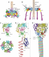
Fig. 1.
Fig. 1.
Structure of the bacteriophage T4 baseplate and comparison of the E. coli CFT073 c3393 VgrG with its T4 homologs, gp5 and gp27. CryoEM reconstructions of the T4 baseplate before (A) and after (B) attachment to the host cell. Component proteins are labeled with their respective gene numbers. The T6SS protein homologs are highlighted in bold and underlined. (C) The crystal structure of the c3393 VgrG. Different domains are colored in distinct colors. The gp27 tube domains are colored cyan and light green. The fragment of the polypeptide chain connecting the gp27 and gp5 modules is shown as a thick red tube. (D) The structures of gp5 and gp27 monomers extracted from the (gp5)3-(gp27)3 complex. The terminal ends of the gp5 and gp27 polypeptide chains, which become fused in the VgrG structure, are highlighted with red dots. (E) A model of the prototypical VgrG is created from the entire (gp5)3-(gp27)3 complex by removing the lysozyme domain. (F) End-on view of the crystal structure of the c3393 VgrG trimer.
Fig. 2.
Fig. 2.
Superposition of the Hcp1 and gp27 structures. (A) Superposition of the crystallographic Hcp1 dimer (red and blue) onto the gp27 monomer (cyan). (B) An end-on view of the superposition of the Hcp1 hexamer (red and blue) onto the entire gp27 trimer (cyan).
Fig. 3.
Fig. 3.
Electron microscopy analysis of oligomerization properties of Hcp proteins. Hcp proteins fused at C terminus to a 3xAla-6xHis-tag were over-expressed in E. coli BL21 Star (Invitrogen) and purified by affinity chromatography using Ni-NTA Agarose (QIAGEN). For electron microscopy, the protein samples were diluted to a final concentration of 0.02 mg/ml and negatively stained with uranyl formate. Electron micrographs were recorded with an FEI Tecnai G2 Spirit BioTWIN electron microscope. Arrows point to polymeric structures. (A) PA1512 (Hcp2, P. aeruginosa PAO1), (B) c3391 (Hcp, E. coli CFT073), (C) PA2367 (Hcp3, P. aeruginosa PAO1), (D) An aliquot of purified PA2367 protein sample was denatured by adding solid urea (Fluka) to 8 M concentration. The protein was refolded by 500× dilution into a buffer without urea.
Fig. 4.
Fig. 4.
ClustalX sequence alignment of T4 gp25 and the T6SS ORF c3402 from E. coli CFT073.
Fig. 5.
Fig. 5.
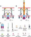
Structure and assembly of the T6SS apparatus. (A and B) Two steps are shown. See text for details. Although the model is based on some reported protein-protein interactions, predicted membrane topologies, and subcellular localization, the majority of the detail presented is speculative. (C) Explanation of color coding and labeling. The proteins, whose names are given in italic, have not been identified in the T6SS cluster yet.

Comment in

References

    1. Economou A, et al. Secretion by numbers: Protein traffic in prokaryotes. Mol Microbiol. 2006;62:308–319. - PMC - PubMed
    1. Pukatzki S, et al. Identification of a conserved bacterial protein secretion system in Vibrio cholerae using the Dictyostelium host model system. Proc Natl Acad Sci USA. 2006;103:1528–1533. - PMC - PubMed
    1. Bingle LE, Bailey CM, Pallen MJ. Type VI secretion: A beginner's guide. Curr Opin Microbiol. 2008;11:3–8. - PubMed
    1. Mougous JD, et al. A virulence locus of Pseudomonas aeruginosa encodes a protein secretion apparatus. Science. 2006;312:1526–1530. - PMC - PubMed
    1. Folkesson A, Lofdahl S, Normark S. The Salmonella enterica subspecies I specific centisome 7 genomic island encodes novel protein families present in bacteria living in close contact with eukaryotic cells. Res Microbiol. 2002;153:537–545. - PubMed

Publication types

MeSH terms

Associated data