. 2009 Mar 24;106(12):4736-41.
doi: 10.1073/pnas.0810053106. Epub 2009 Feb 27.

Noonan syndrome cardiac defects are caused by PTPN11 acting in endocardium to enhance endocardial-mesenchymal transformation

Affiliations

Noonan syndrome cardiac defects are caused by PTPN11 acting in endocardium to enhance endocardial-mesenchymal transformation

Toshiyuki Araki et al. Proc Natl Acad Sci U S A. .

Abstract

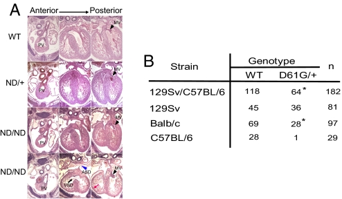
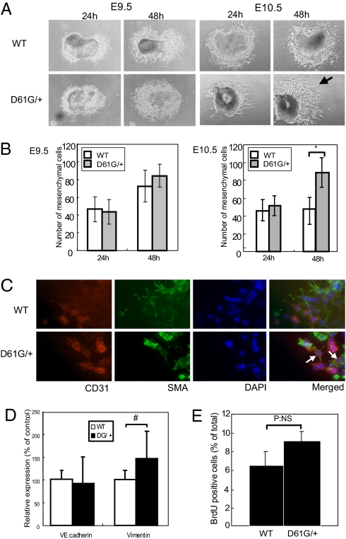
Noonan syndrome (NS), the most common single-gene cause of congenital heart disease, is an autosomal dominant disorder that also features proportionate short stature, facial abnormalities, and an increased risk of myeloproliferative disease. Germline-activating mutations in PTPN11, which encodes the protein tyrosine phosphatase SHP2, cause about half of NS cases; other causative alleles include KRAS, SOS1, and RAF1 mutants. We showed previously that knock-in mice bearing the NS mutant Ptpn11(D61G) on a mixed 129S4/SvJae X C57BL6/J background exhibit all major NS features, including a variety of cardiac defects, with variable penetrance. However, the cellular and molecular mechanisms underlying NS cardiac defects and whether genetic background and/or the specific NS mutation contribute to the NS phenotype remained unclear. Here, using an inducible knock-in approach, we show that all cardiac defects in NS result from mutant Shp2 expression in the endocardium, not in the myocardium or neural crest. Furthermore, the penetrance of NS defects is affected by genetic background and the specific Ptpn11 allele. Finally, ex vivo assays and pharmacological approaches show that NS mutants cause cardiac valve defects by increasing Erk MAPK activation, probably downstream of ErbB family receptor tyrosine kinases, extending the interval during which cardiac endocardial cells undergo endocardial-mesenchymal transformation. Our data provide a mechanistic underpinning for the cardiac defects in this disorder.


Conflict of interest statement

The authors declare no conflict of interest.

Figures

Fig. 1.
Fig. 1.
Mutant Shp2 expression in EC causes NS cardiac defects. (A) Schematic of mutant Ptpn11 loci. Locations of mutations and unique restriction sites are shown. (B) Phosphatase activity of recombinant WT, D61Y, D61G, and N308D Shp2 protein, from Keilhack et al. (12). (C) Sections of hearts from E13.5 WT, ecDY, ncDY, and mcDY embryos. Note the VSD (black arrow), thinned ventricular wall (red arrow), and hypertrophy of pulmonary (blue arrowhead) and AV valves (black arrowhead) observed only in ecDY hearts. (D) Stereologic analysis showing relative valve/cushion volumes of ecDY embryos compared with controls. Values are mean ± SD. n = 3. #, P < 0.05, by 2-tailed student's t test. (E) Efficiency of STOP cassette deletion was analyzed by PCR using outflow tract cushions from WT and ncDY embryos at E10.5 (Top), ventricles from WT and mcDY embryos at E9.5 (Middle) and EC from WT and ecDY at E9.5 (Bottom). The upper band shows the deleted locus; the lower band shows the WT locus.
Fig. 2.
Fig. 2.
Ptpn11 allele and genetic background affect cardiac phenotype. (A) Representative transverse sections of WT, ND/+, and ND/ND embryos. Note the VSD (black arrow), ASD (blue arrowhead), thinned ventricular wall (red arrow), and enlarged AV valves (black arrowheads). The apparent ASD seen in the ND/ND embryo in the third set of panels is not seen in other sections from this embryo. (B) DG/+ mice were backcrossed onto 129Sv or BALB/c for 5 generations or onto C57BL/6 for 3 generations. Viable progeny at 3 weeks are indicated. Note the significantly deceased viability on C57BL/6 background. *, P < 0.05; χ2 test.
Fig. 3.
Fig. 3.
Sustained EMT in D61G/+ EC cushions. (A, B) Explant assays using E9.5 (Left) or E10.5 (Right) EC from WT and DG/+ embryos. Mesenchymal cells were counted at 24 and 48 h. Note that EC from E10.5 DG/+ embryos produce more mesenchymal cells (arrow). Values represent mean ± SD. n = 8. *, P < 0.01, by 2-tailed student's t test. (C) Excess mesenchymal cells are caused by sustained EMT. Cushion explants at E10.5 were stained with the indicated antibodies and DAPI. CD31 and αSMA double-positive “transitional” cells (white arrows) are found only in DG/+ explants. (D) Quantitative RT-PCR of VE-cadherin and vimentin. Values represent mean ± SD. n = 8. #, P < 0.05, by 2-tailed student's t test. (E) Proliferation of cushion mesenchymal cells. Explants (7/genotype) exposed to BrdU for 6 h were immunostained with BrdU antibodies, and BrdU+ cells were counted. NS, not statistically significant.
Fig. 4.
Fig. 4.
D61G mutation enhances EMT by increasing Erk activation. (A, B) Increased Erk activation in DG/+ cushions. EC lysates at E10.5 were immunoblotted with anti-phospho-Erk (Upper) or anti-total Erk2 (Lower) antibodies. Data are quantified in (B). Values are mean ± SD of triplicates. n = 3. #, P < 0.05, by 2-tailed student's t test. (C) Effect of D61G mutant is blocked by Mek inhibitor. U0126 or DMSO as a control was added to explants after 24 h, and mesenchymal cells were counted 24 h later. Values are mean ± SD. n = 8. *. P < 0.01, assessed by 1-way ANOVA; Newman-Keuls multiple range test. (D) Effects of D61G mutation are blocked by ErbB family RTK inhibitors. The indicated inhibitors (PD173074: 60 nM, AG825: 10 μM, AG1478: 100 nM: Tarceva: 300 nM) or DMSO as a control were added to explants after 24 h. Mesenchymal cells were counted 24 h later. Values are mean ± SD. n = 8. Statistical significance (± inhibitor) was assessed by 1-way ANOVA; Newman-Keuls multiple range test (*, P < 0.01).

References

    1. Noonan JA. Noonan syndrome. An update and review for the primary pediatrician. Clinical Pediatrics. 1994;33:548–555. - PubMed
    1. Tartaglia M, Gelb BD. Noonan syndrome and related disorders: Genetics and pathogenesis. Annual Review of Genomics and Human Genetics. 2005;6:45–68. - PubMed
    1. Chan G, Kalaitzidis D, Neel BG. The tyrosine phosphatase Shp2 (PTPN11) in cancer. Cancer Metastasis Rev. 2008;27:179–192. - PubMed
    1. Neel BG, Gu H, Pao L. The ‘Shp’ing news: SH2 domain-containing tyrosine phosphatases in cell signaling. Trends Biochem Sci. 2003;28:284–293. - PubMed
    1. Bentires-Alj M, Kontaridis MI, Neel BG. Stops along the RAS pathway in human genetic disease. Nat Med. 2006;12:283–285. - PubMed

Publication types

MeSH terms

Substances