. 2009 Mar 17;106(11):4085-9.
doi: 10.1073/pnas.0813019106. Epub 2009 Feb 26.

Picomolar sensitivity MRI and photoacoustic imaging of cobalt nanoparticles

Affiliations

Picomolar sensitivity MRI and photoacoustic imaging of cobalt nanoparticles

Louis-S Bouchard et al. Proc Natl Acad Sci U S A. .

Abstract

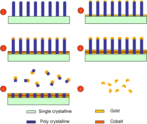
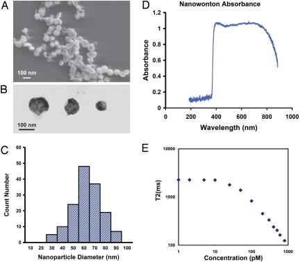
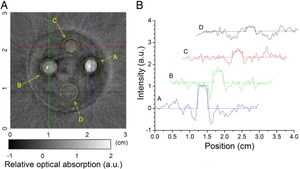
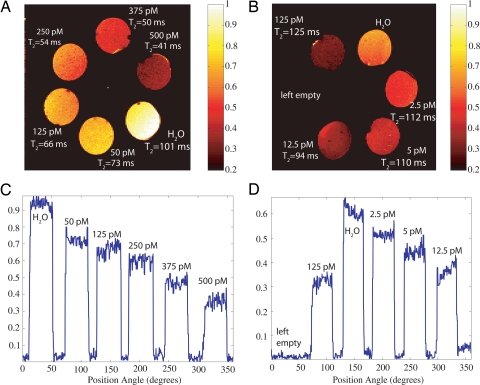
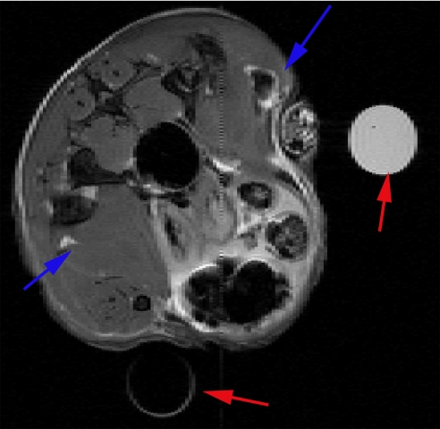
Multimodality imaging based on complementary detection principles has broad clinical applications and promises to improve the accuracy of medical diagnosis. This means that a tracer particle advantageously incorporates multiple functionalities into a single delivery vehicle. In the present work, we explore a unique combination of MRI and photoacoustic tomography (PAT) to detect picomolar concentrations of nanoparticles. The nanoconstruct consists of ferromagnetic (Co) particles coated with gold (Au) for biocompatibility and a unique shape that enables optical absorption over a broad range of frequencies. The end result is a dual-modality probe useful for the detection of trace amounts of nanoparticles in biological tissues, in which MRI provides volume detection, whereas PAT performs edge detection.


Conflict of interest statement

The authors declare no conflict of interest.

Figures

Fig. 1.
Fig. 1.
Fabrication procedure of the nanowontons including 6 steps: (1) Etching polysilicon nanopillars on the surface of single crystalline silicon wafer (for simpler presentation, we omitted from the illustration the preparatory step of depositing 5 nm of chromium before going to step 2, see Methods); (2) deposition of a 10-nm gold thin film; (3) deposition of 10-nm cobalt thin film; (4) deposition of 10-nm gold thin film; (5) etching polysilicon nanopillars in KOH batch solution; and (6) complete removal of polysilicon and chromium by KOH etching and separation of nanowontons.
Fig. 2.
Fig. 2.
Characterization of the cobalt nanowontons. (A) Scanning electron microscopy image of nanowontons. (B) Transmission electron microscopy image of 3 nanowontons in various diameters; notice that the lighter regions in the nanoparticle are relatively hollow, and are responsible for photothermal tuning properties of the nanowonton (38). (C) Particle diameter distribution of 150 nanowontons. Because of the inhomogeneous polysilicon nanopillar diameter, the size of the nanowontons varies from 30 to 90 nm, and the average diameter is 60 nm. (D) Absorption spectrum of nanowonton, medium peak wavelength is ≈700 nm. (E) Spin–spin relaxation time T2 measured at 20 MHz proton frequency and 37 °C. T2 begins to change when the concentration exceeds 20 pM. Note that although 20 MHz is much less than 300 MHz (used for the MRI), this gives a lower bound on relaxivity and shows that the contrast works even at low fields, such as those from portable NMR devices.
Fig. 3.
Fig. 3.
Photoacoustic imaging of nanowonton phantom gels. (A) PAT image of 4 absorbing objects containing nanowonton contrast agent embedded in a gel phantom (5% agarose). The concentrations of nanowontons were 100, 50, 25, and 13 pM, respectively, for objects A, B, C, and D. (B) Intensity profiles extracted from the image along 4 lines (horizontal and vertical dashed lines indicated on the image) going through the absorbing centers are plotted to highlight the visibility of nanowonton inclusions in the reconstructed image. With a CNR close to 1, the object D, where the nanowonton concentration is 13 pM, can hardly be recognized from the background, showing that the current PAT system has detection sensitivity on the order of 25 pM.
Fig. 4.
Fig. 4.
MRI of nanowonton gel phantoms. The nanowonton gels are arranged along the perimeter of a circle. (A and B) Spin-echo images for the phantoms A and B, respectively, with an echo time (TE) of 50 ms and recycle time (TR) of 1 s. The higher-concentration samples appear darker in the images, with doped water used as a control and exhibiting the strongest T2-weighted intensity. The concentrations of the gels are given in the figure along with the T2 values that are deduced from a 7-point curve-fitting procedure. The field of view for this image is 3 × 3 cm, the number of points is 256 × 128, and the slice thickness is 1 mm. (C and D) Intensity profiles for the images in A and B. The relative intensities along a circular contour drawn through the middle of the gels are plotted as a function of the gel azimuthal angle from the x axis. The nanoparticles contrast remains detectable down to 2.5 pM. The images for phantom A and B are plotted to different (normalized) scales.
Fig. 5.
Fig. 5.
Transverse (axial) MRI image in mouse leg muscle injected with Co nanoparticles in PBS solution. The position of the blue arrows indicates the sites of injection for the Co nanoparticles (upper right corner) and the PBS control (lower left corner). Two water-carrying test tubes are also visible in the scans for purposes of MRI slice alignment (red arrows). MRI parameters were: TE = 50 ms; TR = 1 s; field of view is 2.6 cm × 2.6 cm, and slice thickness is 0.5 mm.

References

    1. Al-Jamal W T, Kostarelos K. Liposome-nanoparticle hybrids for multimodal diagnostic and therapeutic applications. Nanomedicine-UK. 2007;2:85–98. - PubMed
    1. Jaffer FA, et al. Cellular imaging of inflammation in atherosclerosis using magnetofluorescent nanomaterials. Mol Imaging. 2006;5:85–92. - PubMed
    1. Mulder WJM, et al. MR molecular imaging and fluorescence microscopy for identification of activated tumor endothelium using a bimodal lipidic nanoparticle. FASEB J. 2005;19 doi: . - DOI - PubMed
    1. Tan WB, Zhang Y. Multi-functional chitosan nanoparticles encapsulating quantum dots and Gd-DTPA as imaging probes for bio-applications. J Nanosci Nanotechno. 2007;7:2389–2393. - PubMed
    1. Dosev D, et al. Magnetic/luminescent core/shell particles synthesized by spray pyrolysis and their application in immunoassays with internal standard. Nanotechnology. 2007;18 055102. - PMC - PubMed

Publication types