. 2009 Mar 17;106(11):4390-5.
doi: 10.1073/pnas.0809080106. Epub 2009 Feb 26.

Apoptosis-stimulating protein of p53 (ASPP2) heterozygous mice are tumor-prone and have attenuated cellular damage-response thresholds

Affiliations

Apoptosis-stimulating protein of p53 (ASPP2) heterozygous mice are tumor-prone and have attenuated cellular damage-response thresholds

Kerstin M Kampa et al. Proc Natl Acad Sci U S A. .

Abstract

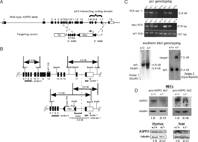
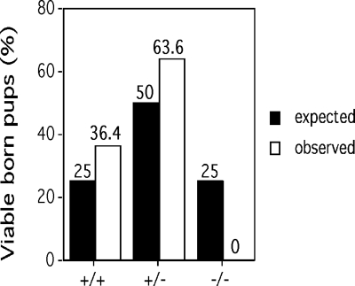
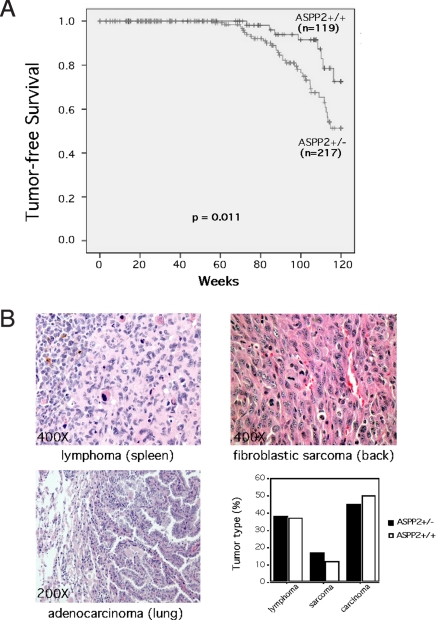
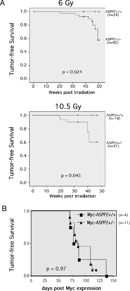
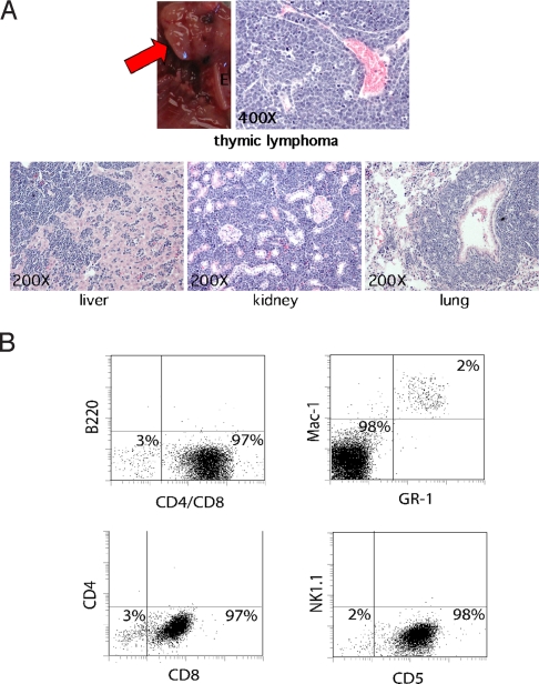
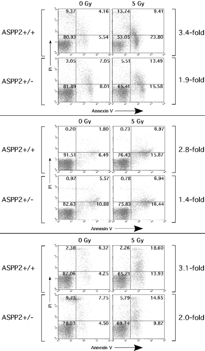
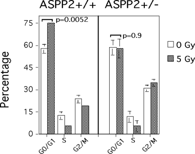
The expression of ASPP2 (53BP2L), a proapoptotic member of a family of p53-binding proteins, is frequently suppressed in many human cancers. Accumulating evidence suggests that ASPP2 inhibits tumor growth; however, the mechanisms by which ASPP2 suppresses tumor formation remain to be clarified. To study this, we targeted the ASPP2 allele in a mouse by replacing exons 10-17 with a neoR gene. ASPP2(-/-) mice were not viable because of an early embryonic lethal event. Although ASPP2(+/-) mice appeared developmentally normal, they displayed an increased incidence of a variety of spontaneous tumors as they aged. Moreover, gamma-irradiated 6-week-old ASPP2(+/-) mice developed an increased incidence of high-grade T cell lymphomas of thymic origin compared with ASPP2(+/+) mice. Primary thymocytes derived from ASPP2(+/-) mice exhibited an attenuated apoptotic response to gamma-irradiation compared with ASPP2(+/+) thymocytes. Additionally, ASPP2(+/-) primary mouse embryonic fibroblasts demonstrated a defective G(0)/G(1) cell cycle checkpoint after gamma-irradiation. Our results demonstrate that ASPP2 is a haploinsufficient tumor suppressor and, importantly, open new avenues for investigation into the mechanisms by which disruption of ASPP2 pathways could play a role in tumorigenesis and response to therapy.


Conflict of interest statement

The authors declare no conflict of interest.

Figures

Fig. 1.
Fig. 1.
Generation of ASPP2+/− mice. (A) Schema of ASPP2 allele and targeting vector. Black boxes indicate coding exons; white boxes indicate untranslated regions. Neo indicates neomycin resistance gene; TK indicates thymidine kinase gene. (B) Strategies for detecting a wild-type ASPP2 allele (Upper) or integration of the targeting vector (Lower). Probe locations for Southern blot analysis are shown as gray bars. Open arrows indicate PCR set 1 primers. Neo-PCR indicates NeoR gene amplicon. WT-PCR indicates ASPP2 exon 13 amplicon. (C) Genotyping using PCR (Upper) and Southern blot analysis (Lower). (D) Western blots on equivalent amounts of total protein from ASPP2+/− and ASPP2+/+ MEFs (Upper), or thymuses and livers (Lower), using anti-ASPP2 Ab1 or anti-ASPP2 Ab2. Fold-expression (relative to +/+) normalized to tubulin.
Fig. 2.
Fig. 2.
ASPP2−/− mice are not viable. Indicated genotypes of newborn pups from ASPP2+/− matings are shown. The expected Mendelian frequencies are indicated by black columns; observed ASPP2 genotypes are indicated by white columns.
Fig. 3.
Fig. 3.
ASPP2+/− mice have an increased incidence of spontaneous tumors. (A) Kaplan–Meier tumor-free survival curves for ASPP2+/+ and ASPP2+/− mice (P = 0.011, log-rank test). (B) H&E-stained microscopic sections of representative tumors found in ASPP2+/− mice and a graph of tumor type frequency between genotypes (P = n.s., Fisher exact test).
Fig. 4.
Fig. 4.
ASPP2+/− mice have an increased incidence of γ-irradiation-induced lymphomas. (A) Kaplan–Meier lymphoma-free survival curves for ASPP2+/+ and ASPP2+/− mice after 6.0-Gy or 10.5-Gy (P = 0.024 or P = 0.045, log-rank test) total γ-irradiation delivered in divided weekly fractions starting at 6 weeks old. (B) Kaplan–Meier lymphoma-free survival curves for tet-o-MYC;EμSR-tTA mice (29) after conditional c-Myc expression in an ASPP2+/+ or ASPP2+/− background (P = 0.97, log-rank test).
Fig. 5.
Fig. 5.
High-grade thymic T cell lymphomas induced in ASPP2+/− mice after γ-irradiation. (A) Thymic lymphoma (red arrow) in an ASPP2+/− mouse. H&E-stained microscopic sections of lymphoma-infiltrated liver, kidney, and lung from this mouse. (B) Immunophenotyping of tumor cells in unfractionated bone marrow from this mouse. A total of 2% of normal myeloid progenitors coexpressed Mac-1 and Gr-1 (Upper Right). A total of 98% of the bone marrow was replaced by tumor cells expressing CD5 and CD8 (Lower), but not B220 (Upper Left), CD4, and Nk1.1 markers (Lower).
Fig. 6.
Fig. 6.
ASPP2+/− thymocytes have an attenuated apoptotic response to γ-irradiation. Flow cytometry on ASPP2+/+ or ASPP2+/− primary thymocytes 4 h after 5-Gy γ-irradiation (right column) or 0-Gy γ-irradiation (left column). The percentage of cells in each quadrant (relative to total cells) is indicated. The fold-increase in apoptosis between 0-Gy and 5-Gy γ-irradiated thymocytes was determined by the increase in percentage of Annexin V-stained cells (lower right plus upper right quadrants).
Fig. 7.
Fig. 7.
ASPP2+/− MEFs have an attenuated G0/G1 checkpoint. Cell cycle distribution shows an increase in G0/G1-arrested ASPP2+/+ MEFs but not ASPP2+/− MEFs 24 h after 5-Gy γ-irradiation (P = 0.0052 and P = 0.9, unpaired 2-tailed Student's t test, respectively). Mean values of triplicate experiments are shown with SDs.

References

    1. Iwabuchi K, Bartel PL, Li B, Marraccino R, Fields S. Two cellular proteins that bind to wild-type but not mutant p53. Proc Natl Acad Sci USA. 1994;91:6098–6102. - PMC - PubMed
    1. Samuels-Lev Y, et al. ASPP proteins specifically stimulate the apoptotic function of p53. Mol Cell. 2001;8:781–794. - PubMed
    1. Takahashi N, et al. Expression of 53BP2 and ASPP2 proteins from TP53BP2 gene by alternative splicing. Biochem Biophys Res Commun. 2004;315:434–438. - PubMed
    1. Lopez CD, et al. Proapoptotic p53-interacting protein 53BP2 is induced by UV irradiation but suppressed by p53. Mol Cell Biol. 2000;20:8018–8025. - PMC - PubMed
    1. Bergamaschi D, et al. ASPP1 and ASPP2: Common activators of p53 family members. Mol Cell Biol. 2004;24:1341–1350. - PMC - PubMed

Publication types

Substances