. 2009 Apr 24;284(17):11326-35.
doi: 10.1074/jbc.M807709200. Epub 2009 Feb 27.

miR-199a, a bone morphogenic protein 2-responsive MicroRNA, regulates chondrogenesis via direct targeting to Smad1

Affiliations

miR-199a, a bone morphogenic protein 2-responsive MicroRNA, regulates chondrogenesis via direct targeting to Smad1

Edward A Lin et al. J Biol Chem. .

Abstract

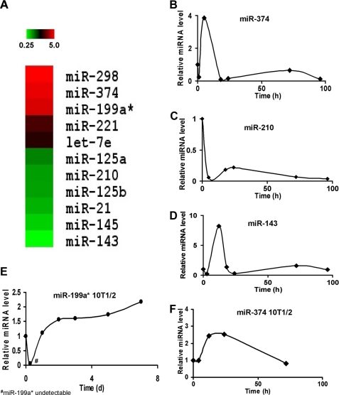
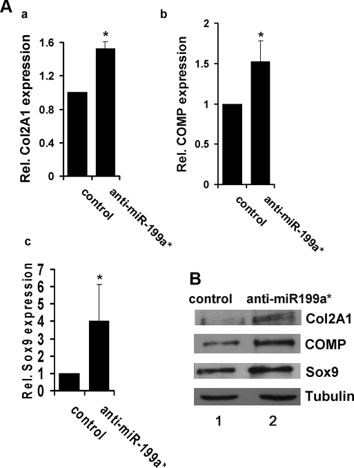
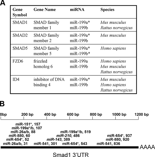
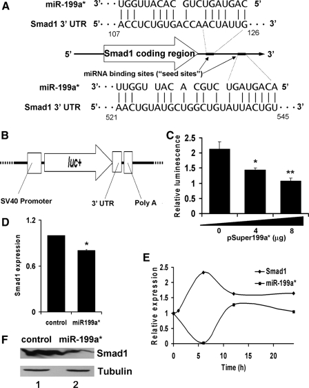
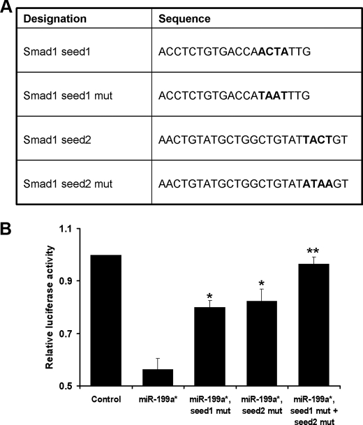
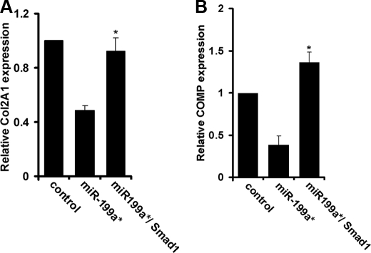
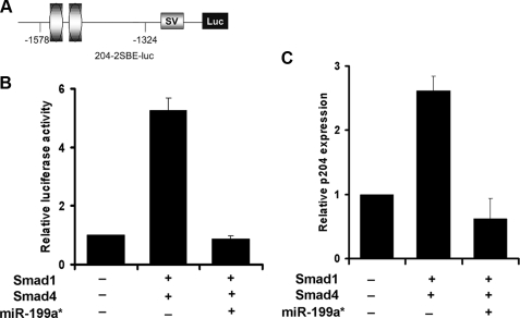
MicroRNAs (miRNA) are short non-coding RNA molecules that regulate a variety of biological processes. The role of miRNAs in BMP2-mediated biological processes is of considerable interest. A comparative miRNA array led to the isolation of several BMP2-responsive miRNAs. Among them, miR-199a(*) is of particular interest, because it was reported to be specifically expressed in the skeletal system. Here we demonstrate that miR-199a(*) is an early responsive target of BMP2: its level was dramatically reduced at 5 h, quickly increased at 24 h and remained higher thereafter in the course of BMP2-triggered chondrogenesis of a micromass culture of pluripotent C3H10T1/2 stem cells. miR-199a(*) significantly inhibited early chondrogenesis, as revealed by the reduced expression of early marker genes for chondrogenesis such as cartilage oligomeric matrix protein (COMP), type II collagen, and Sox9, whereas anti-miR-199a(*) increased the expression of these chondrogenic marker genes. A computer-based prediction algorithm led to the identification of Smad1, a well established downstream molecule of BMP-2 signaling, as a putative target of miR-199a(*). The pattern of Smad1 mRNA expression exhibited the mirror opposite of miR-199a(*) expression following BMP-2 induction. Furthermore, miR-199a(*) demonstrated remarkable inhibition of both endogenous Smad1 as well as a reporter construct bearing the 3-untranslated region of Smad1 mRNA. In addition, mutation of miR-199a(*) binding sites in the 3'-untranslated region of Smad1 mRNA abolished miR-199a(*)-mediated repression of reporter gene activity. Mechanism studies revealed that miR-199a(*) inhibits Smad1/Smad4-mediated transactivation of target genes, and that overexpression of Smad1 completely corrects miR-199a(*)-mediated repression of early chondrogenesis. Taken together, miR-199a(*) is the first BMP2 responsive microRNA found to adversely regulate early chondrocyte differentiation via direct targeting of the Smad1 transcription factor.


Figures

FIGURE 1.
FIGURE 1.
MicroRNA expression following BMP-2 induction. Panel A, expression profile of various micro-RNAs in response to BMP-2 induction. Total microRNA was isolated from BMP2-treated C2C12 cells and hybridized to a microarray chip. The brightest green shading represents a 75% down-regulation in microRNA expression, whereas the brightest red shading represents a 400% up-regulation. The microRNAs are grouped according to expression pattern. Panels BD, real-time PCR measurement of miRNA expression following induction with BMP-2 in C2C12 cells. Relative miRNA expression levels were measured at various time points, as indicated, following BMP-2 treatment of C2C12 cells. Panels E and F, expression profile of miR-199a* and miR-374 following induction with BMP-2 in C3H10T1/2 cells. Relative miRNA expression levels were measured at various time points, as indicated, following BMP-2 treatment of C3H10T1/2 cells.
FIGURE 2.
FIGURE 2.
Overexpression of miR-199a* inhibits chondrogenesis of C3H10T1/2 and ATDC5 cells. Panel A, miR-199a* inhibition of chondrogenic differentiation of a micromass culture of C3H10T1/2 cells. C3H10T1/2 cells were transfected with an empty pSuper vector (control) or pSuper199a*. After 24 h of treatment with BMP-2 (100 ng/ml), micromass cultures were lysed and the expression of chondrogenic differentiation markers, Col2a1 (a) and COMP (b), was measured via quantitative real-time PCR. Data in A and C are the means of three independent cell culture experiments with S.D. indicated. *, p < 0.05. Panel B, overexpression of miR-199a* inhibits chondrogenesis of ATDC5 cells, assayed by immunoblotting. Following transfection with pSuper199a* or control plasmid, ATDC5 cells were maintained in 10 μg/ml human insulin for 3 days, after which cultures were lysed and whole cell lysates were analyzed for expression of Col 2A1, COMP, and Sox9. Tubulin is used as an internal control. Panel C, overexpression of miR-199a* inhibits chondrogenesis of ATDC5 cells, assayed by real-time PCR. ATDC5 cells were processed as described in panel B and mRNA levels of Col2A1 (a), COMP (b), and Sox9 (c) were determined by real-time PCR.
FIGURE 3.
FIGURE 3.
Suppression of miR-199a* enhances chondrogenesis of ATDC5 cells. Panel A, real-time PCR assay. Following transfection with anti-miR199a* plasmids or control plasmid, ATDC5 cells were maintained in 10 μg/ml human insulin for 3 days, after which cultures were lysed for real-time PCR analysis of chondrogenesis markers Col2A1 (a), COMP (b), and Sox9 (c). Data are the means of three independent experiments with S.D. indicated. *, p < 0.05. Panel B, immunoblotting analysis of chondrogenesis markers. ATDC5 cells were processed as described in panel A and protein levels of Col 2A1, COMP, Sox9, and Tubulin (serving as a loading control) were determined by Western blotting.
FIGURE 4.
FIGURE 4.
Predicted targets of differentially regulated miRNA during differentiation. Panel A, putative targets of the miR-199 family. Panel B, diagram of miRNA targeting Smad1 mRNA. Selected miRNA are featured according to their putative binding site in the Smad1 3′-UTR sequence. miRNAs that were up-regulated following C2C12 induction by BMP-2 are bolded. Down-regulated miRNAs are italicized.
FIGURE 5.
FIGURE 5.
Repression of Smad1 by ectopic expression of miR-199a* in C3H10T1/2 cells. Panel A, schematic of miR-199a* inhibition of Smad1 expression. Inhibition of Smad1 occurs via a specific miRNA binding site (seed site) within the 3′-UTR of Smad1 mRNA. The 5′ end of mir-199a* contains a sequence complementary to the seed site. The seed sites are shown in relation to the coding region of Smad1 mRNA. Panel B, schematic of pGL3Smad1 reporter construct. The pGL3Smad1 vector was constructed by inserting a sequence that corresponds to the 3′-UTR of Smad1 mRNA, immediately downstream of the luc reporter of the pGL3-Control vector. Panel C, miR-199a* represses pGL3Smad1 reporter gene activity in a dose-dependent manner. C3H10T1/2 cells were transfected with 1.5 μg of pGL3Smad1 together with 1.5 μg of pSVGal internal control plasmid and varying amounts of pSuper199a* expression plasmid, as indicated. Data are the means of three independent cell culture experiments, with S.D. indicated. *, p < 0.05; **, p < 0.01 when compared with control. Panel D, repression of Smad1 mRNA level by ectopic miR-199a*, assayed by real-time PCR. Cells transfected with the empty pSuper vector alone (control) or with the miR-199a* overexpression plasmid were treated by BMP-2, as indicated. Smad1 mRNA expression was measured by quantitative real-time PCR; the expression level of BMP-2 untreated cells is set to 1. Data are the means of three independent cell culture experiments with S.D. indicated. *, p < 0.05. Panel E, early down-regulation of miR-199a* after BMP-2 exposure correlates with up-regulation of Smad1. C3H10T1/2 cells exposed to BMP-2 were harvested and lysed at various time points, as indicated. Smad1 mRNA and miR-199a* expression were measured via quantitative real-time PCR; the expression level of non-induced cells is set to 1. Panel F, repression of Smad1 protein level by ectopic miR-199a*, assayed by immunoblotting. Following transfection with pSuper199a* or a control plasmid, C3H10T1/2 cells were harvested after 24 h of BMP-2 exposure. Total cell lysates were subjected to immunoblotting analysis with anti-Smad1 or anti-tubulin antibody.
FIGURE 6.
FIGURE 6.
Alteration of one or two seed sites in the 3-UTR of Smad1 mRNA results in a strong reduction or a complete loss of the miR-199a*-mediated repression of Smad1 expression. Panel A, diagrams show the alterations in the first and/or the second of the seed sites in the pGL3Smad1 reporter gene. Panel B, transient transfection assays in C3H10T1/2 cells. The reporter gene specified and the pSVgal internal control plasmid were transfected into C3H10T1/2 cells together with 3 μg of pSuper (CTR) or the pSuper199a* expression plasmid. At 48 h after transfection, the cultures were harvested and the luciferase and β-galactosidase activities were determined. Data are the means of three independent cell culture experiments, with S.D. indicated. *, p < 0.05; **, p < 0.001.
FIGURE 7.
FIGURE 7.
Smad1 overcomes miR-199*-mediated inhibition of chondrocyte differentiation. C3H10T1/2 cells were either transfected with pSuper199a* or co-transfected with pSuper 199a* together with a Smad1 expression plasmid, as indicated. The micromass cultures were stimulated by BMP-2, and the expression of chondrogenic differentiation markers Col2A1 (Panel A) and COMP (Panel B) were assayed by real-time PCR. Means from three independent experiments are shown (error bars indicate S.D.). *, p < 0.05 when compared with cells transfected with pSuper199a* only.
FIGURE 8.
FIGURE 8.
miR-199a* inhibits Smad-dependent transactivation of p204 gene expression. Panel A, schematic of p204-specific reporter construct. The indicated segments from the 5′-flanking region of the p204 gene were linked to an SV40 promoter (SV) and a DNA segment encoding luciferase (Luc). Ovals represent SBEs. Panel B, miR-199a* inhibits Smad-mediated activation of p204-specific reporter genes. The indicated reporter gene construct was transfected into C3H10T1/2 cells together with either the indicated Smad expression plasmids (i.e. Smad1, Smad4) alone or in addition to pSuper199a*, as well as a β-galactosidase internal control plasmid. The luciferase activities were normalized to the β-galactosidase activities. The normalized values were then calibrated against control values, which were arbitrarily set as 1. Panel C, miR-199a* completely abolishes Smad-activated p204 expression in C3H10T1/2 cells. The indicated Smads expression plasmids and pSuper199a* were transfected into C3H10T1/2 cells. Total RNA was extracted and expression of p204 was assayed via real-time PCR. p204 expression was normalized against the endogenous control, glyceraldehyde-3-phosphate dehydrogenase. The normalized values were then calibrated against control values, which were arbitrarily set as 1. Means from three independent experiments are shown (error bars indicate S.D.).

Similar articles

Cited by

References

    1. Lewis, B. P., Burge, C. B., and Bartel, D. P. (2005) Cell 120, 15-20 - PubMed
    1. Chan, S. P., and Slack, F. J. (2006) RNA Biol. 3, 97-100 - PubMed
    1. Chen, C. Z., Li, L., Lodish, H. F., and Bartel, D. P. (2004) Science 303, 83-86 - PubMed
    1. Esau, C., Kang, X., Peralta, E., Hanson, E., Marcusson, E. G., Ravichandran, L. V., Sun, Y., Koo, S., Perera, R. J., Jain, R., Dean, N. M., Freier, S. M., Bennett, C. F., Lollo, B., and Griffey, R. (2004) J. Biol. Chem. 279, 52361-52365 - PubMed
    1. Harris, T. A., Yamakuchi, M., Ferlito, M., Mendell, J. T., and Lowenstein, C. J. (2008) Proc. Natl. Acad. Sci. U. S. A. 105, 1516-1521 - PMC - PubMed

Publication types