. 2009 May 15;18(10):1813-24.
doi: 10.1093/hmg/ddp098. Epub 2009 Feb 27.

Analysis of Ellis van Creveld syndrome gene products: implications for cardiovascular development and disease

Affiliations

Analysis of Ellis van Creveld syndrome gene products: implications for cardiovascular development and disease

Kristen Lipscomb Sund et al. Hum Mol Genet. .

Abstract

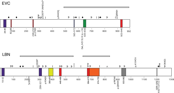
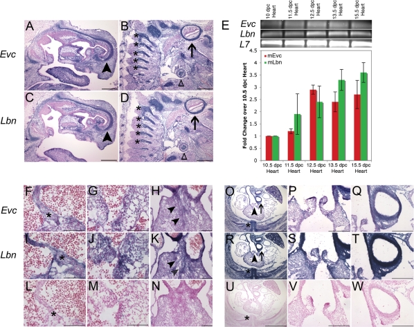
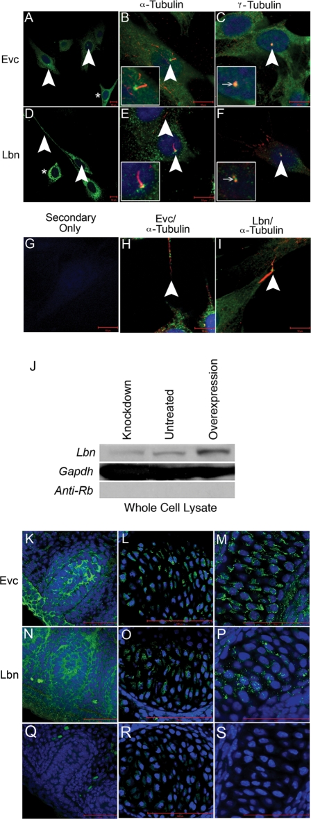
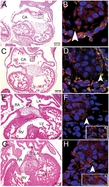
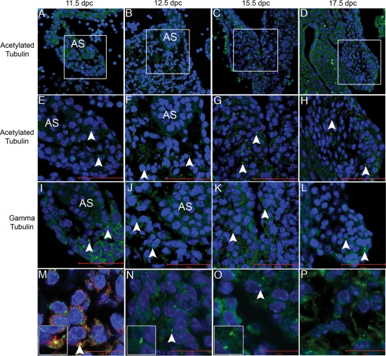
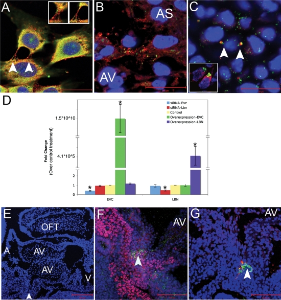
Mutations identified in a cohort of patients with atrioventricular septal defects as a part of Ellis van Creveld syndrome (EvC syndrome) led us to study the role of two non-homologous genes, EVC and LBN, in heart development and disease pathogenesis. To address the cause of locus heterogeneity resulting in an indistinguishable heart-hand phenotype, we carried out in situ hybridization and immunofluorescence and identified co-localization of Evc and Lbn mRNA and protein. In the heart, expression was identified to be strongest in the secondary heart field, including both the outflow tract and the dorsal mesenchymal protrusion, but was also found in mesenchymal structures of the atrial septum and the atrioventricular cushions. Finally, we studied the transcriptional hierarchy of EVC and LBN but did not find any evidence of direct transcriptional interregulation between the two. Due to the locus heterogeneity of human mutations predicted to result in a loss of protein function, a bidirectional genomic organization and overlapping expression patterns, we speculate that these proteins function coordinately in cardiac development and that loss of this coordinate function results in the characteristics of EvC syndrome.


Figures

Figure 1.
Figure 1.
Summary of expected EVC and LBN protein changes based on previously reported mutation data (top of protein) or those identified in this study (bottom of protein) for patients with EvC syndrome. Colored boxes indicate putative domains. Purple, transmembrane; Red, nuclear localization signal; Green, leucine zipper; Blue, ATP/GTP binding site; Yellow, Flagellar hook associated protein; Orange, Helix-loop-helix; Gray, IQ motif; Black, RGD Cell attachment sequence. Shapes indicate predicted protein changes. Diamonds, splicing error; Triangles, insertion or deletion resulting in a frameshift; Delta sign, deletion of a single amino acid; Bar, truncation mutation; Caret, duplication. Deletions are indicated by rectangles over the deleted area. Asterisks denote missense mutations that occur in an amino acid conserved in mouse and chicken. WAD, Weyers Acrodental Dysostosis.
Figure 2.
Figure 2.
Expression of Evc and Lbn mRNA in the developing mouse embryo during AV septation. (AD) Evc and Lbn mRNA expression in a sagittal section of a 15.5 dpc mouse whole embryo. Transcript for both genes is found in the cartilage primordium of the nasal bone (arrowhead), the vertebrae (astericks) and other cartilagenous structures like the primordium of the clavicle (open triangle) and the temporal bone (arrow). The scale bars in (A–D) represent 500 µM. (E) Quantitative RT–PCR confirms the presence of Evc and Lbn message in the developing murine heart from 10 dpc to 15.5 dpc. There is no statistically significant difference in the levels of Evc and Lbn mRNA transcript (P = NS). In situ hybridization in transverse sections of the heart at 13.5 dpc pinpoints co-expression at the tip of the primary atrial septum (asterisks in F and I), in the AV cushions (G and J) and in the connective tissue of the outflow tract (H and K). Sense controls are provided in (LN). Transverse sections at 15.5 dpc reveal Evc and Lbn expression in structures affected in Evc syndrome including the heart (OT) and the ribs (asterisks in O and R). Co-expression in the heart is strongest in outflow tract structures (O–T). The areas pointed out by the arrowhead in (O and R) are magnified in (P and S), while the arrows in (O and R) indicate the areas magnified in (Q and T). Sense controls are provided in (UW). The scale bar represents 200 µM in all images from (F) to (W) except for (Q), (R) and (U), where they represent 1 mm.
Figure 3.
Figure 3.
Novel peptide-specific EVC antibodies confirm localization of Evc protein and co-staining with the newly generated LBN antibody reveals these proteins are expressed in overlapping structures. Expression of EVC alone (A) and co-staining with acetylated tubulin (B) or gamma tubulin (C) in NIH3T3 cells confirms antibody specificity by identification of EVC at the base of cilia. Expression of LBN alone (D) and co-staining with acetylated tubulin (E) or gamma tubulin (F) reveals an overlap of EVC and LBN protein in the cilia. Asterisks in (A and D) show protein presence in the cytoplasm of the cell outside of cilia structures. (G) serves as a negative control while (H and I) reveal the presence of EVC and LBN overlapping with acetylated tubulin-based structures that are not cilia. All scalebars in (AI) represent 10 µM. Western blot for LBN protein (J) identified a band of the estimated size, 148 kDa. Compared with untreated cultures, the intensity of the LBN band in whole cell lysates decreased when treated with Lbn siRNA (Knockdown) and increased upon transfection with Sport6-pCMV-mLbn (Overexpression). GAPDH is used as a loading control and anti-Rb secondary antibody is a negative control for the western blot protocol. These findings confirm LBN antibody specificity. EVC antibody specificity is further confirmed in previously reported in vivo structures including the vibrissae (K), the vertebrae (L) and the cartilage primordium of the nasal bone (M) in a 15.5 dpc sagittal section of an embryo. LBN immunofluorescence reveals overlapping expression in these structures (NP). Negative controls (QS) show minimal background. All scalebars in images (KS) represent 50 µM.
Figure 4.
Figure 4.
EVC and LBN protein colocalize at the tip of the primary atrial septum during murine cardiac development. Development of the atrial septum at stages 10.5–13.5 dpc (A,C,E and G). (B,D,F and H) are higher magnification images of the boxed area in the left column. The primary atrial septum shows colocalization of EVC (Red) and LBN (Green) at the tip of the developing septum as it dives down from the common atrial wall (10.5 and 11.5 dpc) and merges with the AV cushions (12.5 and 13.5 dpc). After the atrial septum joins the endocardial cushions, co-expression broadens to these AV structures (inset, F). The inset box in (H) represents overlap of EVC and LBN proteins along the edge of the atrial septum (location indicated by asterisk in G). Arrowheads are used to pinpoint areas of colocalization at the tip of the atrial septum. Blue color indicates nuclear staining with Topro-3. Scalebars represent 200 µM in (A,C,E and G). CA, common atrium; RA, right atrium; LA, left atrium; R, right ventricle; LV, left ventricle.
Figure 5.
Figure 5.
Acetylated tubulin, a marker of cilia that colocalizes with EVC in cell culture, is present in the developing heart. Acetylated tubulin (seen here in green) is identified in the primary atrial septum (A,E and I), the AV junction (B,F and J) and the developing AV valves (C,D,G,H,K and L). Boxes in (A,B,C and D) indicate areas of higher magnification in (E–L). (M) displays co-staining of both acetylated and gamma tubulin confirming the presence of cilia. In images (E–N), the scale bars depict 50 µM while the scale bar represents 20 µM in (M–P). (N) is a higher magnification of acetylated tubulin revealing the cilia axonene and (O) is a higher magnification of gamma tubulin, which marks the basal bodies. (P) shows evidence of acetylated tubulin outside the cilia in mesenchymal structures of the heart. Blue Topro-3 staining marks the nuclei. Arrowheads indicate individual cilia.
Figure 6.
Figure 6.
EVC and LBN proteins are coexpressed in cell culture and in cells crucial for cardiac AV septation but there is no hierarchal transcriptional interregulation. EVC and LBN proteins are coexpressed in a similar pattern in NIH3T3 cells (A) and in vivo at the tip of the atrial septum (B) of a 12.5 dpc mouse. Arrows indicate overlapping expression of EVC and LBN in bone (C) and in the developing AV valve at 13.5 dpc (inset in C). Knockdown and overexpression of EvC-associated genes reveals a lack of hierarchial transcriptional interregulation (D). Manipulation of gene expression in cell culture revealed that changes in expression of EVC did not impact the levels of Lbn transcript and changes in the expression of LBN did not affect Evc transcription. The blue bars indicate successful knockdown of Evc after treatment with siRNA, but no change in Lbn gene expression. The red bars show Lbn could be knockeddown but the treatment did not affect Evc transcription. The yellow bar represents transfection with a control plasmid or a control siRNA. The green bar indicates successful overexpression of EVC with no affect on levels of Lbn transcription. The purple bar shows transfection of LBN does not alter the levels of Evc in the cell. Asterisks indicate statistical significance based on the Bonferroni adjusted P-value <0.0125. (EG) are sagittal sections of a mouse heart at 10.5 dpc. AS, atrial septum; AV, atrioventricular cushions; A, atria; V, ventricles; OFT, outflow tract. The arrowhead in (E) indicates the area of zoom in (F) and (G). (F) shows overlap of ISL-1 (pink) and LBN (green) while G shows overlap of EVC (red) and LBN (green) in the DMP.

References

    1. Ellis R.W.B., van Creveld S. A syndrome characterized by ectodermal dysplasia, polydactyly, chondro-dysplasia and congenital morbus cordis: report of three cases. Arch. Dis. Child. 1940;15:65–84. - PMC - PubMed
    1. McKusick V.A., Egeland J.A., Eldridge R., Krusen D.E. Dwarfism in the Amish I.The Ellis-van Creveld syndrome. Bull. Johns Hopkins Hosp. 1964;115:306–336. - PubMed
    1. McKusick V.A. Ellis-van Creveld syndrome and the Amish. Nat. Genet. 2000;24:203–204. - PubMed
    1. Ruiz-Perez V.L., Ide S.E., Strom T.M., Lorenz B., Wilson D., Woods K., King L., Francomano C., Freisinger P., Spranger S., et al. Mutations in a new gene in Ellis-van Creveld syndrome and Weyers acrodental dysostosis. Nat. Genet. 2000;24:283–286. - PubMed
    1. Galdzicka M., Patnala S., Hirshman M.G., Cai J.F., Nitowsky H., Egeland J.A., Ginns E.I. A new gene, EVC2, is mutated in Ellis-van Creveld syndrome. Mol. Genet. Metab. 2002;77:291–295. - PubMed

Publication types

MeSH terms