. 2009 May;75(9):2861-8.
doi: 10.1128/AEM.01317-08. Epub 2009 Feb 27.

DNA as an adhesin: Bacillus cereus requires extracellular DNA to form biofilms

Affiliations

DNA as an adhesin: Bacillus cereus requires extracellular DNA to form biofilms

Sébastien Vilain et al. Appl Environ Microbiol. 2009 May.

Abstract

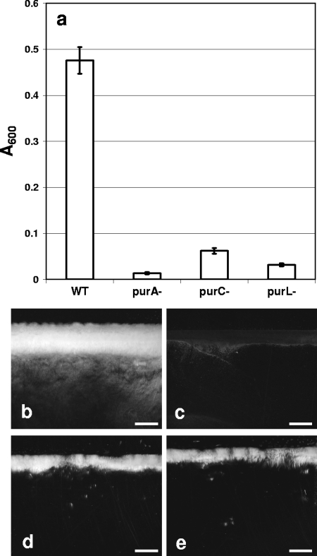
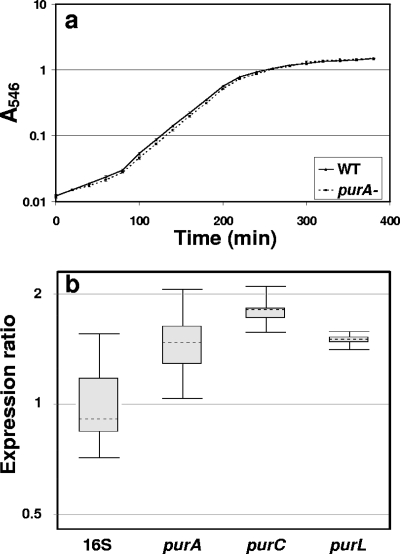
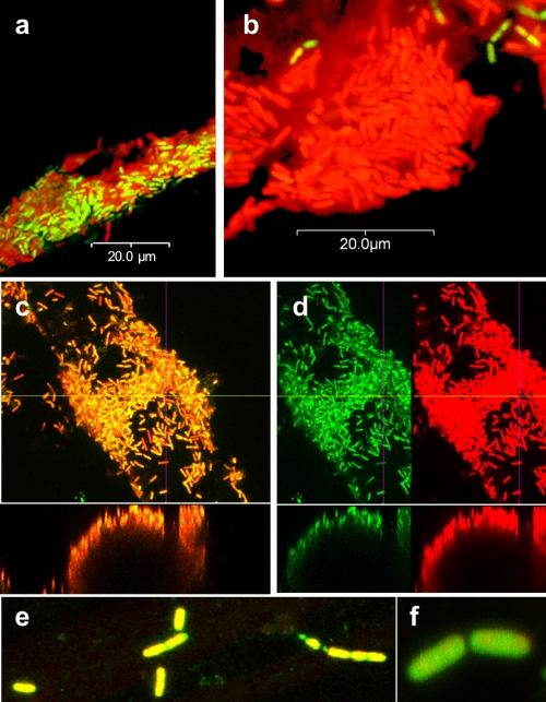
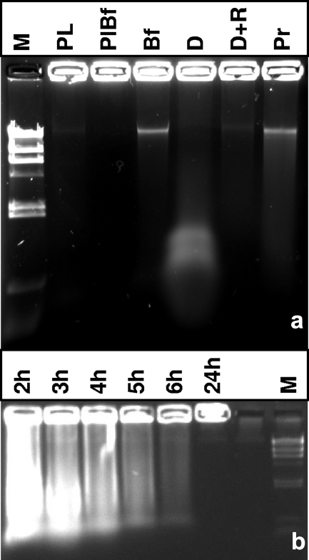
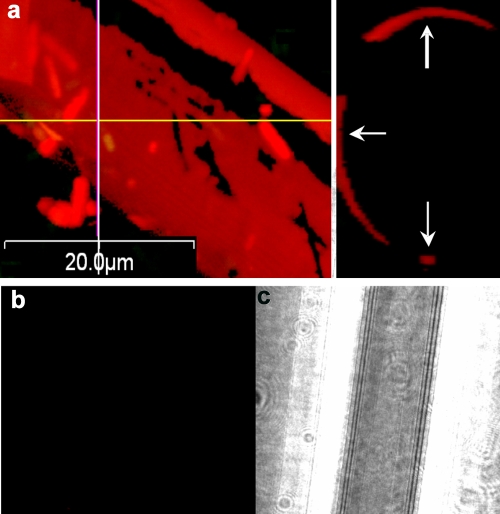
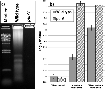
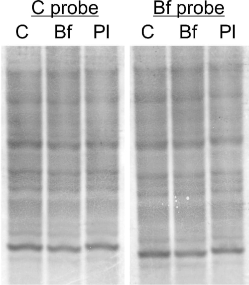
The soil saprophyte Bacillus cereus forms biofilms at solid-liquid interfaces. The composition of the extracellular polymeric matrix is not known, but biofilms of other bacteria are encased in polysaccharides, protein, and also extracellular DNA (eDNA). A Tn917 screen for strains impaired in biofilm formation at a solid-liquid interface yielded several mutants. Three mutants deficient in the purine biosynthesis genes purA, purC, and purL were biofilm impaired, but they grew planktonically like the wild type in Luria-Bertani broth. Biofilm populations had higher purA, purC, and purL transcript ratios than planktonic cultures, as measured by real-time PCR. Laser scanning confocal microscopy (LSCM) of BacLight-stained samples indicated that there were nucleic acids in the cell-associated matrix. This eDNA could be mobilized off the biofilm into an agarose gel matrix through electrophoresis, and it was a substrate for DNase. Glass surfaces exposed to exponentially growing populations acquired a DNA-containing conditioning film, as indicated by LSCM. Planktonic exponential-phase cells released DNA into an agarose gel matrix through electrophoresis, while stationary-phase populations did not do this. DNase treatment of planktonic exponential-phase populations rendered cells more susceptible than control populations to the DNA-interacting antibiotic actinomycin D. Exponential-phase purA cells did not contain detectable eDNA, nor did they convey a DNA-containing conditioning film to the glass surface. These results indicate that exponential-phase cells of B. cereus ATCC 14579 are decorated with eDNA and that biofilm formation requires DNA as part of the extracellular polymeric matrix.


Figures

FIG. 1.
FIG. 1.
Biofilm formation by B. cereus ATCC 14579 and selected transposon mutants. (a) Biofilm formation was quantified by dislodging the biomass from the walls of glass beakers following static incubation in LB broth at 25°C for 72 h. The error bars indicate the standard deviations of the means (P < 0.01, n = 5). WT, wild type. (b to e) Stereophotomicrographs of biofilms of the wild type (b) and purA (c), purC (d), and purL (e) mutants on glass slides at the air-liquid interface. Scale bars = 2 mm.
FIG. 2.
FIG. 2.
(a) Growth of the wild-type and purA mutant strains in LB broth at 28°C. (b) Box plot indicating the ratio of the pur gene transcripts in a biofilm to the pur gene transcripts in planktonic populations quantified by real-time PCR using the 16S rRNA gene as an endogenous control. WT, wild type. 16S, 16S rRNA gene.
FIG. 3.
FIG. 3.
LSCM of a 24-h BacLight-stained B. cereus biofilm cultured on glass wool at 28°C for 24 h. (a and b) Some sections of the biofilm contained cells that appeared to be both red and green (a), while the majority of the cells appeared to be red and to be surrounded by a less dense red area (propidium iodide fluorescence) (b). (c and d) Cells that appeared to be red (c) did contain green-fluorescing centers (Syto 9) when they were viewed in separate channels (d). Images for green and red channels set at identical detection values are shown separately, and images of x-z cross sections are shown below the large images. (e and f) The purA mutant cells formed very sparse biofilms and did not have an apparent red exterior.
FIG. 4.
FIG. 4.
Biofilms and exponential-phase planktonic cells of B. cereus contain eDNA. (a) A biofilm population (lane Bf), a planktonic population (lane Pl), and a planktonic stationary-phase population from a biofilm culture flask (lane PlBf) were placed in wells of a 0.8% agarose gel, along with biofilm treated with DNase (lane D), with RNase plus DNase (lane D+R), and with proteinase K (lane P). Lane M contained HindIII-digested λ DNA. (b) Ethidium bromide-stained agarose gel containing planktonic populations from cultures that were different ages. Populations were harvested by centrifugation after they were cultured in LB broth for 2, 3, 4, 5, 6, and 24 h and normalized for cell density, and cell suspensions were loaded directly into the wells for electrophoresis. Lane M contained HindIII-digested λ DNA as a size marker.
FIG. 5.
FIG. 5.
(a) Glass wool exposed for 30 min to an exponentially growing B. cereus culture before removal and staining with propidium iodide, viewed by LSCM. The left panel is a composite x-y image, and the right panel is a y-z image of the in silico preparation at the position of the line. (b and c) Glass wool exposed to sterile LB broth for 2 h and then stained with propidium iodide and viewed by LSCM in the red channel (b) and by differential interference contrast microscopy (c).
FIG. 6.
FIG. 6.
(a) The purA mutant does not appear to be decorated with eDNA like the wild type. The presence of eDNA was determined electrophoretically by placing 20-μl portions of washed exponential-phase suspensions (optical density at 546 nm, 5.0) of wild-type and purA mutant populations in a 0.8% agarose gel, which was resolved by electrophoresis at 60 V for 60 min. The marker was HindIII-digested λ DNA. (b) Susceptibility of exponential-phase wild-type and purA mutant populations to actinomycin D determined with and without prior treatment with DNase. Mid-exponential-phase planktonic cultures were washed and treated either with 5 U/ml RNase-free DNase or with water for 30 min prior to exposure to 50 μg·ml−1 actinomycin D.
FIG. 7.
FIG. 7.
Southern hybridization of biofilm eDNA (lane Bf), exponential-phase planktonic eDNA (lane Pl), and chromosomal DNA (lane C). Biofilm and planktonic eDNA were obtained by electrophoresis from 24-h biofilm and 2-h planktonic populations, followed by extraction from the gel. DNA was digested with EcoRI, and all three extracts were hybridized to digoxigenin-labeled chromosomal DNA (C probe) or biofilm eDNA (Bf probe).

References

    1. Aguilar, C., H. Vlamakis, R. Losick, and R. Kolter. 2007. Thinking about Bacillus subtilis as a multicellular organism. Curr. Opin. Microbiol. 10:638-643. - PMC - PubMed
    1. Allesen-Holm, M., K. B. Barken, L. Yang, M. Klausen, J. S. Webb, S. Kjelleberg, S. Molin, M. Givskov, and T. Tolker-Nielsen. 2006. A characterization of DNA release in Pseudomonas aeruginosa cultures and biofilms. Mol. Microbiol. 59:1114-1128. - PubMed
    1. Belem, M. A. F., B. F. Gibbs, and B. H. Lee. 1997. Enzymatic production of ribonucleotides from autolysates of Kluyveromyces marxianus grown on whey. J. Food Sci. 62:851-854+857.
    1. Böckelmann, U., A. Janke, R. Kuhn, T. R. Neu, J. Wecke, J. R. Lawrence, and U. Szewzyk. 2006. Bacterial extracellular DNA forming a defined network-like structure. FEMS Microbiol. Lett. 262:31-38. - PubMed
    1. Bone, E. J., and D. J. Ellar. 1989. Transformation of Bacillus thuringiensis by electroporation. FEMS Microbiol. Lett. 49:171-177. - PubMed

Publication types

LinkOut - more resources