. 2009 Nov;41(5):583-9.
doi: 10.1165/rcmb.2008-0201OC. Epub 2009 Feb 27.

WNT5A is a regulator of fibroblast proliferation and resistance to apoptosis

Affiliations

WNT5A is a regulator of fibroblast proliferation and resistance to apoptosis

Louis J Vuga et al. Am J Respir Cell Mol Biol. 2009 Nov.

Abstract

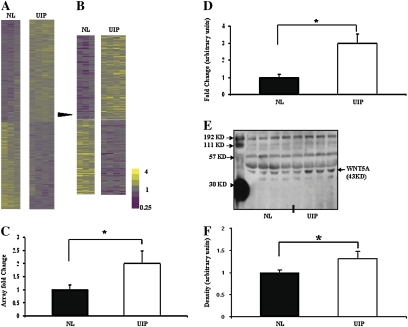
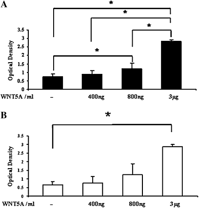
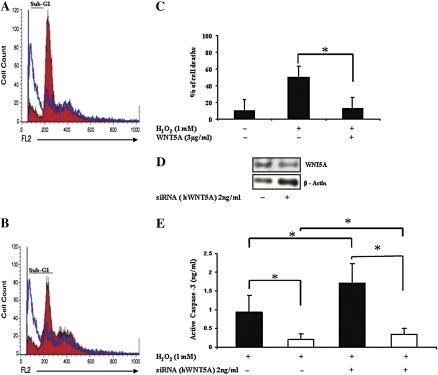
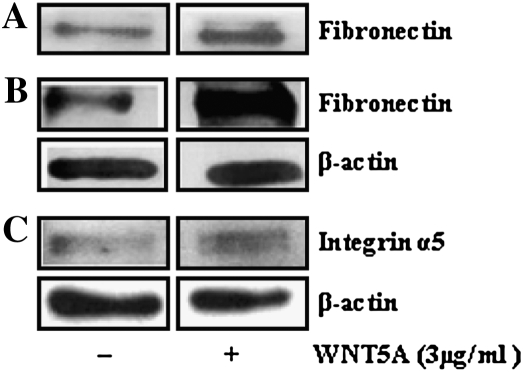
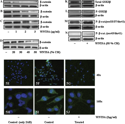
Usual interstitial pneumonia (UIP) is a specific histopathologic pattern of interstitial lung fibrosis that may be idiopathic or secondary to autoimmune diseases and environmental exposures. In this study, we compared gene expression patterns in primary fibroblasts isolated from lung tissues with UIP histology and fibroblasts isolated from lung tissues with normal histology using expression microarrays. We found that WNT5A was significantly increased in fibroblasts obtained from UIP lung tissues compared with normal lung fibroblasts, an observation verified by quantitative real-time RT-PCR and Western blot. Because the role of WNT5A in UIP is unknown, we treated normal lung fibroblasts or UIP lung fibroblasts with WNT5A, and found that WNT5A increased proliferation as well as relative resistance to H2O2-induced apoptosis. This effect was not mediated through the canonical WNT/beta-catenin pathway, as WNT5A induced a decrease in beta-catenin levels in the same cells. In addition, WNT5A induced increases in fibronectin and alpha(5)-integrin in normal lung fibroblasts. Collectively, our data suggest that WNT5A may play a role in fibroblast expansion and survival characteristics of idiopathic pulmonary fibrosis and other fibrotic interstitial lung diseases that exhibit UIP histological patterns.


Figures

Figure 1.
Figure 1.
WNT5A is overexpressed in usual interstitial pneumonia (UIP) lung fibroblasts. (A) Heat map of 8,396 genes that had an Entrez Gene ID demonstrated differences in gene expression patterns between UIP lung fibroblast lines (n = 4) and normal lung (NL) fibroblast lines (n = 3). (B) A total of 350 genes substantially changed (TNoM = 0 and t test, P < 0.05). Up-regulated genes are presented in yellow, down-regulated genes in purple, and unchanged genes in gray. (C) WNT5A expression levels in microarray data. WNT5A gene expression was increased in UIP compared with NL (TNoM = 0, t test, P < 0.05). (D) Quantitative RT-PCR (qRT-PCR) verification of microarray data demonstrate increased WNT5A in UIP (n = 5) compared with NL (n = 5) (P < 0.05). (E) WNT5A protein levels were higher in UIP (n = 4) than NL (n = 4), as measured by Western blot analysis of cell supernatant. The loading control is the band at 192 kD. (F) Densitometric quantification of WNT5A protein levels measured by Western analysis in E. WNT5A was significantly higher in UIP than NL (*P < 0.05). All fibroblast lines were used at early passages (–7).
Figure 2.
Figure 2.
WNT5A promotes fibroblasts proliferation. (A) NL fibroblasts (solid bars; n = 4) and (B) UIP lung fibroblasts (open bars; n = 3) were treated with 400 ng/ml, 800 ng/ml, or 3 μg/ml of WNT5A, cultured for 72 hours, and optical densities, as a measure of cell proliferations, were significantly increased in a dose-dependant manner (*P < 0.05). Note that UIP lung fibroblasts were less responsive to WNT5A when compared with control (P values < 0.05). The figures shown are representatives of three experiments.
Figure 3.
Figure 3.
WNT5A inhibits H2O2-induced apoptosis. NL fibroblast lines (n = 3) were pretreated with WNT5A or vehicle control for 4 hours before H2O2 treatment. (A) Comparison of apoptosis as determined by cell count in the sub-G1 fraction between control (red) and 1 mM H2O2-treated cells (blue). Cells treated with 1 mM H2O2 showed more apoptotic cells in the sub-G1 fraction than control cells. (B) Comparison between combined treatments. Pretreatment of cells with WNT5A before the addition of 1 mM H2O2 (red) reduced the number of apoptotic cells compared with 1 mM H2O2-treated cells (blue). (C) Means of apoptotic cells in the sub-G1 fractions of three experiments expressed by a bar for each treatment condition. WNT5A pretreatment inhibited H2O2-induced fibroblast apoptosis (*P < 0.05). (D) Transfection of NL fibroblasts with WNT5A siRNA (hWNT5A) decreases protein expression of WNT5A 72 hours after transfection. (E) Silencing WNT5A using siRNA (hWNT5A) induced active caspase-3 in NL fibroblast lines (solid bars; n = 3) and UIP lung fibroblast lines (open bars; n = 3). The concentration of active caspase-3 was significantly increased (*P < 0.05) in cells treated with both 1 mM H2O2 and 2 ng/ml siRNA (hWNT5A) compared with control cells (scrambled siRNA) treated with H2O2. Active caspase-3 levels were lower in UIP fibroblasts compared with control cells.
Figure 4.
Figure 4.
WNT5A effects on protein expression. (AC) NL fibroblast lines treated with 3 μg/ml WNT5A showed an increase in fibronectin levels both in supernatant (A) and lysates (B). (C) WNT5A-treated NL showed higher level of α5-integrin than untreated NL. β-Actin was used as a loading control.
Figure 5.
Figure 5.
WNT5A decreases β-catenin levels. (A) NL fibroblasts, (B) UIP lung fibroblasts, and (C) A549 cells treated with 1, 2, or 3 μg/ml WNT5A showed a dose-dependent reduction of β-catenin protein level. (D) Treatment of NL with conditioned media (CM) containing WNT5A. Using Western blot analysis, a decreased level of β-catenin was observed (upper panel) and equal loading of protein was verified by applying anti–β-actin (lower panel). (EJ) Level of β-catenin visualized using immunofluorescence microscope. Cells were stained with anti–β-catenin (green) and DNA counterstaining, blue. Although a bright green staining can be observed in control cells (F and I), this staining intensity is reduced after treatment with 3 μg/ml WNT5A (G and J), similar to the background staining of negative control cells (E and H). (KL) WNT signaling in lung fibroblasts. WNT5A (Supernatant) showed no effect on total GSK3β levels and P- GSK3β (ser9) levels, and no change in P–β-catenin (ser33/37/thr41) level in treated NL (KM). (N) The protein level of phospho–β-catenin (ser45/thr41) showed an increase in WNT5A-treated NL compared with control cells. Anti–β-actin served as standard control for equal loading of each protein. All images were cut from the same individual gels.

Comment in

References

    1. Lynch J III, Saggar R, Weigt S, Zisman D, White E. Usual interstitial pneumonia. Semin Respir Crit Care Med 2006;27:634–651. - PubMed
    1. Katzenstein A, Myers J. Idiopathic pulmonary fibrosis: clinical relevance of pathologic classification. Am J Respir Crit Care Med 1998;157:1301–1315. - PubMed
    1. Meltzer E, Noble P. Idiopathic pulmonary fibrosis. Orphanet J Rare Dis [serial on the Internet]. 2008;3:8. Available from: http://www.ojrd.com/content/3/1/8 - PMC - PubMed
    1. Katzenstein A, Zisman D, Litzky L, Nguyen B, Kotloff R. Usual interstial pneumonia histologic study of biopsy and explant specimens. Am J Surg Pathol 2002;26:1567–1577. - PubMed
    1. Selman M, King TE Jr, Pardo A. Idiopathic pulmonary fibrosis: prevailing and evolving hypotheses about its pathogenesis and implications for therapy. Ann Intern Med 2001;134:136–151. - PubMed

Publication types

MeSH terms