. 2009 Feb;21(2):460-78.
doi: 10.1105/tpc.108.064444. Epub 2009 Feb 27.

The RPN5 subunit of the 26s proteasome is essential for gametogenesis, sporophyte development, and complex assembly in Arabidopsis

Affiliations

The RPN5 subunit of the 26s proteasome is essential for gametogenesis, sporophyte development, and complex assembly in Arabidopsis

Adam J Book et al. Plant Cell. 2009 Feb.

Abstract

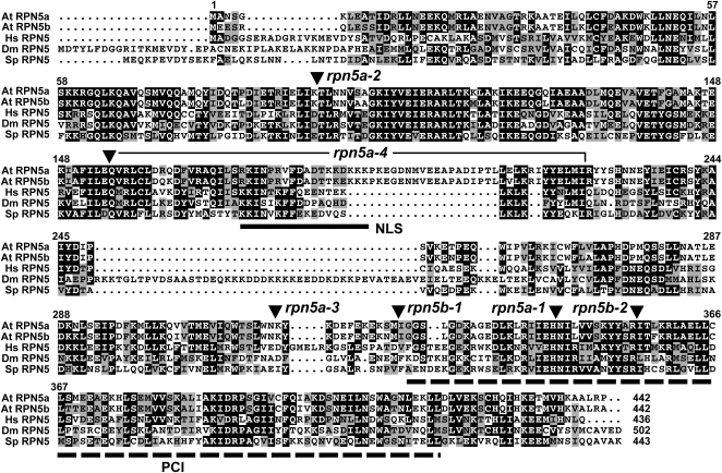
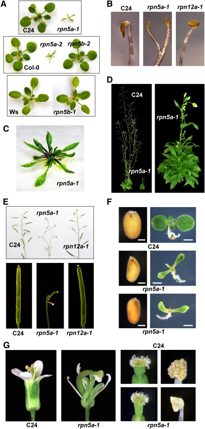
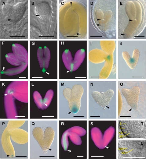
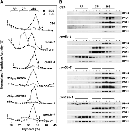
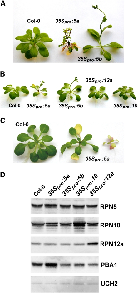
The 26S proteasome is an essential multicatalytic protease complex that degrades a wide range of intracellular proteins, especially those modified with ubiquitin. Arabidopsis thaliana and other plants use pairs of genes to encode most of the core subunits, with both of the isoforms often incorporated into the mature complex. Here, we show that the gene pair encoding the regulatory particle non-ATPase subunit (RPN5) has a unique role in proteasome function and Arabidopsis development. Homozygous rpn5a rpn5b mutants could not be generated due to a defect in male gametogenesis. While single rpn5b mutants appear wild-type, single rpn5a mutants display a host of morphogenic defects, including abnormal embryogenesis, partially deetiolated development in the dark, a severely dwarfed phenotype when grown in the light, and infertility. Proteasome complexes missing RPN5a are less stable in vitro, suggesting that some of the rpn5a defects are caused by altered complex integrity. The rpn5a phenotype could be rescued by expression of either RPN5a or RPN5b, indicating functional redundancy. However, abnormal phenotypes generated by overexpression implied that paralog-specific functions also exist. Collectively, the data point to a specific role for RPN5 in the plant 26S proteasome and suggest that its two paralogous genes in Arabidopsis have both redundant and unique roles in development.


Figures

Figure 1.
Figure 1.
Amino Acid Sequence Comparison of RPN5 Proteins. Identical and similar residues are shown in the black and gray boxes, respectively. Dots denote gaps. At, Arabidopsis thaliana; Hs, Homo sapiens; Dm, Drosophila melanogaster; Sp, Schizosaccharomyces pombe. The numbers refer to the amino acid position in At RPN5a. The residue length of each protein is shown at the end of the sequence. The locations of the PCI and NLS sequences are indicated by the dashed and solid lines, respectively. The positions of the various insertion mutants are indicated by the arrowheads. The bracket highlights the coding region that has been deleted in the rpn5a-4 allele.
Figure 2.
Figure 2.
Expression Patterns of Arabidopsis RPN5a and RPN5b Genes and Proteins. (A) Relative transcript abundance in various tissues determined from the GENEVESTIGATOR DNA microarray data set (Zimmermann et al., 2004). Sus, suspension culture; Yg, young; Mat, mature; Sen, senescent. Error bars represent the se from different arrays. (B) Expression of RPN5apro:GUS (5a) and RPN5bpro:GUS (5b) in 12-d-old seedlings (bars = 2 mm), floral tissue (bars = 25 μm), root tips (bars = 100 μm), trichome socket cells (bars = 200 μm), and developing embryos (bars = 25 μm). Tissues were stained overnight in X-Gluc. (C) Subcellular localization of RPN5a-GFP and RPN5b-GFP in the roots of rpn5a-1 seedlings as detected by confocal fluorescence microscopy (bars = 50 μm). The middle panels show a fourfold magnification of cells (bars = 10 μm). The panels below demonstrate the rescue of the rpn5a-1 mutant phenotype by the 35Spro:RPN5a-GFP and 35Spro:RPN5b-GFP transgenes (bars = 2 mm).
Figure 3.
Figure 3.
Molecular and Biochemical Descriptions of Arabidopsis rpn5a and rpn5b Mutants. (A) Organization of the RPN5a and RPN5b genes and the positions of the T-DNA insertions. The bracket for rpn5a-4 defines the deleted region. White and black boxes indicate untranslated and coding regions, respectively. Lines denote introns. (B) RNA gel blot analysis of 2-week-old wild-type seedlings, homozygous mutant seedlings, and wild-type seedlings expressing 35Spro:RPN5a or 35Spro:RPN5b transgenes. Equal loading of total RNA was confirmed by probing the blots with β-tubulin4 (TUB4). (C) RNA gel blot analysis of RPN5a and RPN5b transcripts in 2-week-old rpn10-1, rpn12a-1, and rpt2a-2 mutant seedlings. (D) Immunoblot detection of 26S proteasome subunits and Ub. Equivalent amounts of total protein (20 μg) were subjected to SDS-PAGE and immunoblot analysis with antibodies against RPN5a, RPN12a, PBA1, and Ub. Equal protein loads were confirmed by probing with anti-UBC1 antibodies. The positions of free Ub, poly-Ub chains, and Ub conjugates are indicated.
Figure 4.
Figure 4.
Phenotype of rpn5a Mutants. (A) Stunted growth of 2-week-old rpn5a seedlings compared with wild-type and rpn5b seedlings grown under continuous light. Each mutant is compared with its corresponding wild-type parent: C24 for rpn5a-1, Col-0 for rpn5a-2 and rpn5b-2, and Ws for rpn5b-1. (B) Premature apical hook opening and increased petiole elongation of etiolated rpn5a seedlings compared with wild-type and rpn12a-1 seedlings. Seedlings were grown for 6 d in darkness. (C) Stunted growth of 5-week-old rpn5a-1 seedling. (D) Dwarfed influorescence of a 2-month-old rpn5a-1 plant compared with wild-type C24. (E) Infertility of the rpn5a-1 mutant. Top panel, inflorescence of mature flowering plants. Bottom panel, close-up of developing siliques. (F) Abnormal size and shape of hydrated rpn5a-1 seeds compared with a wild-type C24 sibling. Left panels, seeds before germination. Right panels, resulting 10-d-old seedlings. Bars = 0.1 mm (seeds) and 1 mm (seedlings). (G) Abnormal floral development of rpn5a-1 plants. Left panels, whole flowers. Right panels, close-ups of the stigma and anthers from mature flowers.
Figure 5.
Figure 5.
Phenotype and Expression Patterns of Wild-Type and Homozygous rpn5a Embryos. For each set of embryos, the wild type and mutant are shown at approximately the same developmental age. In all cases, the wild-type embryos are from heterozygous siliques of the same line as the corresponding mutant. In the fluorescent microscopy images, the tissue is highlighted by chlorophyll autofluorescence (false-colored magenta). In (A) and (B), arrowheads point to the abnormal shape of the cells in the position of the QC in rpn5a-1. In (C) to (U), arrowheads locate the RAM and SAM in the wild-type embryos or the equivalent position in the mutants. C, cortex; E, endodermis; QC?, cells in the QC position; C/E?, single cell file of unknown identity in the position of the cortex/endodermis. Bars = 50 μm. (A) and (B) Young embryos of the wild type (A) and the rpn5a-4 mutant (B) at the heart stage. (C) Mature embryos of the wild type and the rpn5a mutants. (D) Example of the short embryo phenotype (rpn5a-2). (E) Example of a long embryo phenotype (rpn5a-1). (F) to (H) DR5rev:GFP expression in the wild type at the bent cotyledon stage (F) and rpn5a-4 mutant embryos with normal expression (G) and high expression that extended apically (H). (I) and (J) STMpro:GUS expression in the wild type at the bent cotyledon stage (I) and rpn5a-4 (J). (K) and (L) CUC1 (M0233 line) expression in the wild type at the bent cotyledon stage (K) and rpn5a-4 (L). (M) to (O) PIN4pro:GUS expression in the wild type at the torpedo stage (M) and rpn5a-4 with no expression (N) or weak expression (O). (P) and (Q) QC25pro:GUS expression in the wild type at the bent cotyledon stage (P) and rpn5a-4 (Q). (R) and (S) SCRpro:GFP expression in the wild type at the bent cotyledon stage (R) and rpn5a-4 (S). (T) and (U) Close-up of the root meristem in the wild type at the late heart stage (T) and rpn5a-3 (U) embryos.
Figure 6.
Figure 6.
Instability of the 26S Proteasome from the rpn5a-1 Mutant. 26S proteasomes were enriched from 10-d-old liquid-grown seedlings of the indicated genotypes and subjected to centrifugation in a 10 to 40% glycerol gradient. The general location of the 26S proteasome and the 20S CP and 19S RP subcomplexes are indicated by the brackets. (A) Assay of gradient fractions for CP peptidase activity in the absence or presence of 0.02% SDS using the substrate Suc-LLVY-AMC. The activity scale for each profile was adjusted to generate a near equal height for the peak activity. The arrows delineate the range of fractions that were used for the immunoblot analysis (B). (B) Immunoblot analysis of gradient fractions for the presence of various 26S proteasome subunits.
Figure 7.
Figure 7.
Rescue of the rpn5a Mutant Phenotype with RPN5a and RPN5b. rpn5a-1 mutants in the C24 background were transformed with the RPN5a or RPN5b genomic coding regions expressed under the control of the CaMV 35S promoter (35Spro), the RPN5a promoter (5apro), or the RPN5b promoter (5bpro). (A) Genotyping of complementation lines. DNA was isolated from 10-d-old seedlings and PCR amplified with primers specific for wild-type RPN5a gene, the T-DNA in rpn5a-1 (rpn5a-1 T-DNA), or the RPN5a and RPN5b transgenes (trans). Plants homozygous (homo) and heterozygous (het) for the rpn5a-1 T-DNA insertion were included for comparison. (B) Levels of RPN5 protein in the complementation lines. Crude protein extracts were prepared from 10-d-old seedlings and subjected to SDS-PAGE and immunoblot analysis with antibodies against RPN5a, RPN1a, RPN10, RPN12a, and PBA1. Analysis with anti-HSP70 antibodies was used to confirm equal protein loads. (C) Phenotypic rescue of rpn5a-1 mutants. Pictures of 14-d-old plants homozygous for the rpn5a-1 mutation and homozygous for the RPN5a or RPN5b transgenes.
Figure 8.
Figure 8.
Effects of Overexpression of RPN5a and RPN5b on Arabidopsis Development. The RPN5a and RPN5b cDNAs were expressed under the control of the CaMV 35S promoter in wild-type Col-0 seedlings. mRNA levels are shown in Figure 2B. (A) Phenotype of 3-week-old 35Spro:RPN5a and 35Spro:RPN5b plants grown in continuous light. (B) Early flowering of 35Spro:RPN5a and 35Spro:RPN5b plants grown in continuous light compared with Col-0 seedlings overexpressing two other RP subunits (35Spro:RPN10 and 35Spro:RPN12a). (C) Premature leaf senescence of 35Spro:RPN5a plants grown in continuous light. Pictured are two independent lines with differing degrees of severity next to wild-type Col-0. (D) Levels of the corresponding RP proteins in the RPN5a-, RPN5b-, RPN10-, and RPN12a-overexpressing plants shown in (A). Crude protein extracts were prepared from 7-d-old green plants and subjected to immunoblot analysis with antibodies against RPN5a, RPN10, RPN12a, and PBA1. Analysis with anti-UCH1 antibodies was used to confirm equal protein loads.
Figure 9.
Figure 9.
Acceleration of Flowering Time by Exposure of Seedlings to MG132. (A) Wild-type Col-0 plants grown without or with 80 μM MG132 for 4 weeks under a short-day photoperiod. (B) and (C) Leaf number at the onset of flowering for seedlings grown under a short-day photoperiod (B) or under continuous light (C) with or without exposure to various concentrations of MG132. Each bar represents the average (±sd) of 10 seedlings.

References

    1. Aida, M., Beis, D., Heidstra, R., Willemsen, V., Blilou, I., Galinha, C., Nussaume, L., Noh, Y.S., Amasino, R., and Scheres, B. (2004). The PLETHORA genes mediate patterning of the Arabidopsis root stem cell niche. Cell 119 109–120. - PubMed
    1. Babiychuk, E., Fuangthong, M., Van Montagu, M., Inze, D., and Kushnir, S. (1997). Efficient gene tagging in Arabidopsis thaliana using a gene trap approach. Proc. Natl. Acad. Sci. USA 94 12722–12727. - PMC - PubMed
    1. Bailly, E., and Reed, S.I. (1999). Functional characterization of RPN3 uncovers a distinct 19S proteasomal subunit requirement for ubiquitin-dependent proteolysis of cell cycle regulatory proteins in budding yeast. Mol. Cell. Biol. 19 6872–6890. - PMC - PubMed
    1. Brukhin, V., Gheyselinck, J., Gagliardini, V., Genschik, P., and Grossniklaus, U. (2005). The RPN1 subunit of the 26S proteasome in Arabidopsis is essential for embryogenesis. Plant Cell 17 2723–2737. - PMC - PubMed
    1. Cary, A.J., Che, P., and Howell, S.H. (2002). Developmental events and shoot apical meristem gene expression patterns during shoot development in Arabidopsis thaliana. Plant J. 32 867–877. - PubMed

Publication types

MeSH terms

Substances