. 2009 Mar;84(3):289-97.
doi: 10.4065/84.3.289.

Ventricular tachycardia and sudden cardiac death

Affiliations

Ventricular tachycardia and sudden cardiac death

Bruce A Koplan et al. Mayo Clin Proc. 2009 Mar.

Erratum in

  • Mayo Clin Proc. 2009 May;84(5):483

Abstract

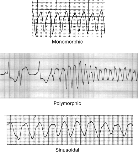
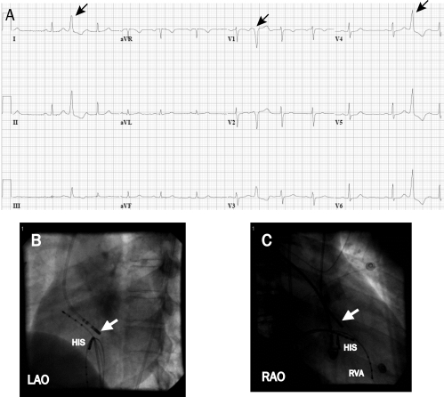
Ventricular tachycardia (VT), which most commonly occurs in patients with structural heart disease, can be associated with an increased risk of sudden death. The most common cause of ventricular fibrillation is acute coronary ischemia, whereas a myocardial scar from prior infarct is the most common cause of sustained monomorphic VT in patients with structural heart disease. More benign forms of idiopathic VT can also occur in the absence of structural heart disease. Treatment of VT involves both emergent management and prevention of recurrence with medical and device therapy. Appropriately selected patients who have experienced VT or those who are at risk of VT may be candidates for an implantable cardioverter-defibrillator. The left ventricular ejection fraction is most frequently used to stratify patients with either ischemic or nonischemic cardiomyopathy who are at risk of sudden death and may be candidates for a prophylactic defibrillator. Catheter ablation may also be an option for appropriately selected patients with many forms of VT. This article discusses the etiologies and management of VT and its association with sudden death.


Figures

FIGURE 1.
FIGURE 1.
Electrograms showing 3 different types of ventricular tachycardia.
FIGURE 2.
FIGURE 2.
Emergent management of hemodynamically unstable ventricular tachycardia (VT)/ventricular fibrillation (VF). Fab = fragment antigen binding compound; MVT = monomorphic VT; NaHCO3 = bicarbonate of soda; PMVT= polymorphic VT. Adapted from the American Heart Association Guidelines for Cardiopulmonary Resuscitation and Emergency Cardiovascular Care.
FIGURE 3.
FIGURE 3.
Twenty-month mortality rate in patients with ischemic heart disease and left ventricular ejection fraction of ≤30% from the second Multicenter Automatic Defibrillator Implantation Trial (MADIT-II). CI = confidence interval; HR = hazard ratio; ICD = implantable cardioverter-defibrillator.
FIGURE 4.
FIGURE 4.
Five-year mortality rate in patients with cardiomyopathy of either ischemic or nonischemic etiology from the Sudden Cardiac Death in Heart Failure Trial (SCD-HeFT). CI = confidence interval; HR = hazard ratio; ICD = implantable cardioverter-defibrillator.
FIGURE 5.
FIGURE 5.
Electrograms showing premature ventricular contractions originating from the aortic annulus. This patient had severely symptomatic premature ventricular contractions and nonsustained ventricular tachycardia of the same morphology. A, The premature ventricular contractions (arrows) have a left bundle branch block configuration, indicating an origin in the right ventricle or septal aspect of the left ventricle. The frontal plane axis is directed inferiorly (dominant R wave in leads II and III). This morphology is consistent with an origin in the outflow region of the left or right ventricle. B and C, Successful site of ablation in the aortic annulus on fluoroscopy. The focus was found at the aortic annulus above the His bundle region (HIS) (white arrow at ablation catheter that enters the ventricle from a retrograde aortic approach). LAO = left anterior oblique; RAO = right anterior oblique; RVA = right ventricular apex.
FIGURE 6.
FIGURE 6.
Voltage map of the left ventricle from a patient with a prior anterior wall infarction viewed from the left lateral and inferior aspect. Colors represent the bipolar electrogram amplitude. Purple represents normal amplitude (>1.5 mV). Electrogram amplitude is progressively lower as colors progress from blue to green to yellow to red. Gray areas are electrically unexcitable, indicating dense fibrosis. Ventricular tachycardia circuits are located in the scar area. For ablation of ventricular tachycardia, additional mapping is required to define the location of the reentry circuit within the scar.

Similar articles

Cited by

References

    1. Coronel R, Fiolet JW, Wilms-Schopman JG, Opthof T, Schaapherder AF, Janse MJ. Distribution of extracellular potassium and electrophysiologic changes during two-stage coronary ligation in the isolated, perfused canine heart. Circulation 1989;80(1):165-177 - PubMed
    1. Pogwizd SM, Corr PB. Mechanisms underlying the development of ventricular fibrillation during early myocardial ischemia. Circ Res. 1990;66(3):672-695 - PubMed
    1. Wit AL, Allessie MA, Bonke FI, Lammers W, Smeets J, Fenoglio JJ., Jr Electrophysiologic mapping to determine the mechanism of experimental ventricular tachycardia initiated by premature impulses: experimental approach and initial results demonstrating reentrant excitation. Am J Cardiol. 1982;49(1):166-185 - PubMed
    1. El-Sherif N, Smith RA, Evans K. Canine ventricular arrhythmias in the late myocardial infarction period, 8: epicardial mapping of reentrant circuits. Circ Res. 1981;49(1):255-265 - PubMed
    1. Lloyd EA, Zipes DP, Heger JJ, Prystowsky EN. Sustained ventricular tachycardia due to bundle branch reentry. Am Heart J. 1982;104(5, pt 1):1095-1097 - PubMed

MeSH terms

Substances