. 2009;4(3):e4658.
doi: 10.1371/journal.pone.0004658. Epub 2009 Mar 2.

Structural basis and catalytic mechanism for the dual functional endo-beta-N-acetylglucosaminidase A

Affiliations

Structural basis and catalytic mechanism for the dual functional endo-beta-N-acetylglucosaminidase A

Jie Yin et al. PLoS One. 2009.

Abstract

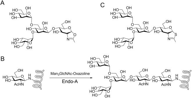
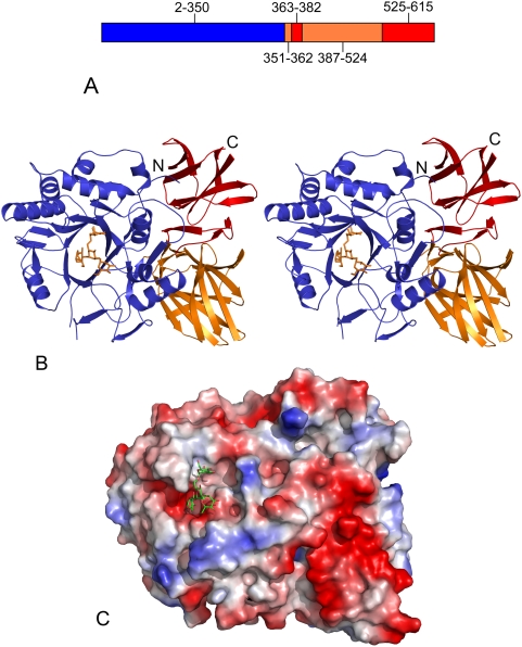
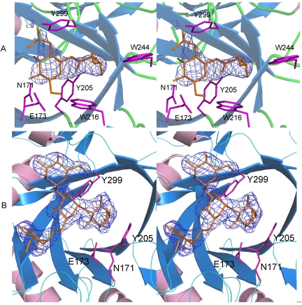
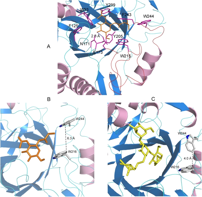
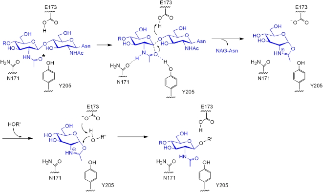
Endo-beta-N-acetylglucosaminidases (ENGases) are dual specificity enzymes with an ability to catalyze hydrolysis and transglycosylation reactions. Recently, these enzymes have become the focus of intense research because of their potential for synthesis of glycopeptides. We have determined the 3D structures of an ENGase from Arthrobacter protophormiae (Endo-A) in 3 forms, one in native form, one in complex with Man(3)GlcNAc-thiazoline and another in complex with GlcNAc-Asn. The carbohydrate moiety sits above the TIM-barrel in a cleft region surrounded by aromatic residues. The conserved essential catalytic residues - E173, N171 and Y205 are within hydrogen bonding distance of the substrate. W216 and W244 regulate access to the active site during transglycosylation by serving as "gate-keepers". Interestingly, Y299F mutation resulted in a 3 fold increase in the transglycosylation activity. The structure provides insights into the catalytic mechanism of GH85 family of glycoside hydrolases at molecular level and could assist rational engineering of ENGases.


Conflict of interest statement

Competing Interests: The authors have declared that no competing interests exist.

Figures

Figure 1
Figure 1. Synthesis of glycopeptides using oligosaccharide oxazolines as sugar donors.
(A) Mimic of oxazoline ion intermediate, Man3GlcNAc-oxazoline. (B) Endo-A catalyzed glycopeptide synthesis by using Man3GlcNAc-oxazoline. (C) An Endo-A inhibitor, Man3GlcNAc-thiazoline.
Figure 2
Figure 2. Structure of Endo-A.
(A) Diagrammatic representation of Endo-A. Amino acids 1–350 make up Domain 1 (blue), segments 351–362 and 387–524 make up Domain 2 (orange), while segments 363–386 and 525–611 make up Domain 3 (red). (B) Stereo image of Endo-A. Man3GlcNAc-thiazoline moiety is shown as sticks. (C) A surface electrostatic potential representation of Endo-A showing the Man3GlcNAc-thiazoline moiety sitting inside the active site cleft.
Figure 3
Figure 3. Stereo images of Endo-A in complex with GlcNAc-Asn (panel A) and Man3GlcNAc-thiazoline (panel B).
The ligand omitted |Fo|-|Fc| αcalc electron density map, calculated using a 3.5 Å resolution data set, shown in blue, was contoured at 2.5σ.
Figure 4
Figure 4. Active site of Endo-A.
(A) N171, E173 and Y299 are critical for catalysis. Amino acids surrounding the carbohydrate moiety are shown as sticks. The distances are shown in dashed lines. The GlcNAc-Asn moiety is shown as sticks. (B) W216 and W244 are “gate-keeping” the active site by sterically regulating access to the active site by the acceptor. The side chain of W244 moves during transglycosylation; the distance between the two Trp is much wider and allows passage of an acceptor into the active site. Trp and GlcNAc-Asn are shown as sticks. (C) The gate is “closed” in the structure of free protein (not shown) and Man3GlcNAc-thiazoline bound Endo-A (shown as sticks).
Figure 5
Figure 5. Transglycosylation activity of Endo-A mutants.
Time course of transglycosylation reaction of wildtype Endo-A (squares) and the three mutants, N171A (circles), Y205F (triangles), and Y299F (diamonds). The reactions were carried out using a chitinase-coupled assay, in which Man9GlcNAc2Asn-Fmoc was used as the donor substrate and 4MU-GlcNAc as the acceptor, 0.25 µg of each enzyme was used in a 50 µl system.
Figure 6
Figure 6. Mechanism of Endo-A mediated catalysis.
(A) Substrate surrounded by critical amino acids - N171, E173 and Y205. The nucleophilic oxygen (O) is marked with *. (B) Intramolecular nucleophilic attack. (C) Formation of an oxazoline ion intermediate. (D) Second nucleophilic attack on the intermediate. (E) Synthesis of a new glycosidic bond. R' could be a GlcNAc molecule of an acceptor during transglycosylation.

References

    1. Suzuki T, Yan Q, Lennarz WJ. Complex, two-way traffic of molecules across the membrane of the endoplasmic reticulum. J Biol Chem. 1998;273:10083–10086. - PubMed
    1. Takegawa K, Nakoshi M, Iwahara S, Yamamoto K, Tochikura T. Induction and purification of endo-beta -N-acetylglucosaminidase from Arthrobacter protophormiae grown in ovalbumin. Appl Environ Microbiol. 1989;55:3107–3112. - PMC - PubMed
    1. Bourgerie S, Karamanos Y, Grard T, Julien R. Purification and characterization of an endo-N-acetyl-beta -D-glucosaminidase from the culture medium of Stigmatella aurantiaca DW4. J Bacteriol. 1994;176:6170–6174. - PMC - PubMed
    1. Fujita K, Takami H, Yamamoto K, Takegawa K. Characterization of endo-beta -N-acetylglucosaminidase from alkaliphilic Bacillus halodurans C-125. Biosci, Biotechnol, Biochem. 2004;68:1059–1066. - PubMed
    1. Kadowaki S, Yamamoto K, Fujisaki M, Kumagai H, Tochikura T. A novel endo-beta -N-acetylglucosaminidase acting on complex oligosaccharides of glycoproteins in a fungus. Agric Biol Chem. 1988;52:2387–2389. - PubMed

Publication types

MeSH terms

Substances