. 2009;4(3):e4663.
doi: 10.1371/journal.pone.0004663. Epub 2009 Mar 2.

Suppression of radiation-induced salivary gland dysfunction by IGF-1

Affiliations

Suppression of radiation-induced salivary gland dysfunction by IGF-1

Kirsten H Limesand et al. PLoS One. 2009.

Abstract

Background: Radiation is a primary or secondary therapeutic modality for treatment of head and neck cancer. A common side effect of irradiation to the neck and neck region is xerostomia caused by salivary gland dysfunction. Approximately 40,000 new cases of xerostomia result from radiation treatment in the United States each year. The ensuing salivary gland hypofunction results in significant morbidity and diminishes the effectiveness of anti-cancer therapies as well as the quality of life for these patients. Previous studies in a rat model have shown no correlation between induction of apoptosis in the salivary gland and either the immediate or chronic decrease in salivary function following gamma-radiation treatment.

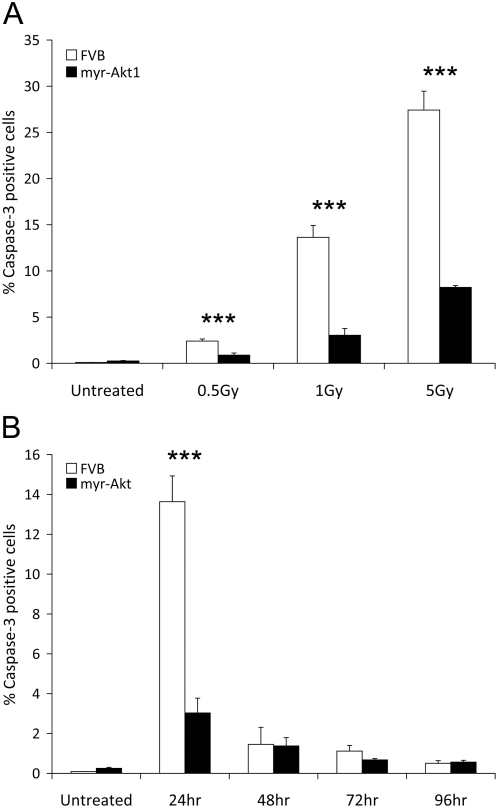
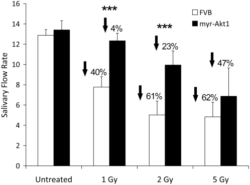
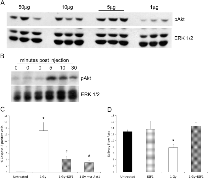
Methodology/principal finding: A significant level of apoptosis can be detected in the salivary glands of FVB mice following gamma-radiation treatment of the head and neck and this apoptosis is suppressed in transgenic mice expressing an activated mutant of Akt (myr-Akt1). Importantly, this suppression of apoptosis in myr-Akt1 mice preserves salivary function, as measured by saliva output, three and thirty days after gamma-radiation treatment. In order to translate these studies into a preclinal model we found that intravenous injection of IGF1 stimulated activation of endogenous Akt in the salivary glands in vivo. A single injection of IGF1 prior to exposure to gamma-radiation diminishes salivary acinar cell apoptosis and completely preserves salivary gland function three and thirty days following irradiation.

Conclusions/significance: These studies suggest that apoptosis of salivary acinar cells underlies salivary gland hypofunction occurring secondary to radiation of the head and neck region. Targeted delivery of IGF1 to the salivary gland of patients receiving head and neck irradiation may be useful in reducing or eliminating xerostomia and restoring quality of life to these patients.


Conflict of interest statement

Competing Interests: The authors have declared that no competing interests exist.

Figures

Figure 1
Figure 1. Reduced apoptosis in myr-Akt1 parotid glands following exposure to ionizing radiation.
In A, four-week old female FVB and myr-Akt1 mice were exposed to 0.5, 1, or 5 Gy γ-radiation and parotid salivary glands were removed 24 hours post-irradiation. Tissues were embedded into paraffin and sections were stained for activated caspase-3. The number of caspase-3 positive cells is graphed as a percentage of the total number of cells per field of view. In B, four-week old female FVB and myr-Akt1 mice were exposed to 1 Gy γ-irradiation and parotid salivary glands were removed at 24, 48, 72 and 96 hours. Tissues were processed for activated caspase-3 immunohistochemistry as described in A. Five fields of view were quantitated for each tissue section and graphed using the averages and SEM from three mice per group. Significant differences (p≤0.05) were determined using a two sample t-test comparing FVB to myr-Akt1 and significant differences are marked with an asterisk (*).
Figure 2
Figure 2. Preservation of salivary flow rates 3 days after single γ-radiation exposure in myr-Akt1 mice.
Four-week old female FVB and myr-Akt1 mice were exposed to 1, 2, or 5 Gy γ-radiation. Three days after irradiation total saliva was collected (over a five minute period) following carbachol injection. Statistical analysis was performed using Student's t-test in Microsoft Excel. Results shown are from ten mice per group in the 1 and 2 Gy doses and four mice per group in the 5 Gy dose. Graphs represent averages and SEM from all mice. Significant differences (p≤0.05) were determined using a two sample t-test comparing FVB to myr-Akt1 and significant differences are marked with an asterisk (*).
Figure 3
Figure 3. IGF1 activates Akt in vivo and preserves salivary flow rates 3 days after single γ-radiation exposure.
In A, FVB mice received an injection of 1, 5, 10, or 50 µg recombinant IGF1. Tissue lysates were collected for immunoblotting five minutes after injection and membranes were probed for activation of Akt using a phosphorylation specific antibody. Results shown are representative of three independent experiments. In B, FVB mice received an injection of 5 µg recombinant IGF1 and tissue lysates were collected for immunoblotting 0, 5, 10 or 30 minutes after injection. Membranes were probed for activated Akt as described in A. Membranes were stripped and re-probed with a total antibody against ERK1/2 as a loading control in both A and B. Results shown are representative of three independent experiments. In C, four-week old female FVB mice were injected with 5 µg recombinant IGF1 immediately prior to treatment with 1 Gy γ-radiation. Salivary glands were removed 24 hours post-irradiation and stained for activated caspase-3 as described in Figure 1A. Graph represents averages and SEM from at least three mice/treatment. (*) indicates significant difference (p≤0.05) from untreated FVB and (#) indicates significance between 1 Gy FVB and 1 Gy IGF1 or 1 Gy myr-Akt1. In D, four-week old female mice were injected with 5 µg recombinant IGF1 and treated with radiation as described in C. Total saliva was collected following carbachol injection 3 days after radiation exposure as described in Figure 2. Statistical analysis was performed using Student's t-test in Microsoft Excel. Results shown are from ten irradiated FVB mice and eight IGF1 plus irradiation mice and graphed using the averages and SEM from all mice. Significant differences (p≤0.05) were determined using a two sample t-test comparing FVB to myr-Akt1 and significant differences are marked with an asterisk (*).
Figure 4
Figure 4. Preservation of salivary flow rates 30 days after single γ-radiation exposure by myr-Akt1 or injection with IGF1.
Four-week old female FVB, myr-Akt1 and FVB mice injected with 5 µg recombinant IGF1 were exposed to 1 Gy γ-radiation. IGF1 injections were performed immediately prior to radiation exposure as described in Figure 3. Thirty days after exposure to γ-radiation total saliva was collected following carbachol injection as described in Figure 2. Statistical analysis was performed using multiple comparison testing in the SAS system. Results shown are from ten irradiated FVB mice, ten irradiated myr-Akt1 mice and eight IGF1 plus irradiation mice and graphed using the averages and SEM from all mice. Treatment groups with the same letters are not significantly different from each other.
Figure 5
Figure 5. Histological analysis of salivary glands from normal and irradiated salivary glands from untreated and IGF1-treated mice.
A) Normal submandibular salivary gland showing basic structure of acini (arrows) and ducts (asterisk). B) Normal sublingual salivary gland. C) Normal parotid gland. D) Submandibular salivary gland thirty days following exposure to 5 Gy radiation. Note area of focal fibrosis and associated inflammatory cells (asterisk). E) Sublingual salivary gland after thirty days following exposure to 5 Gy radiation. No significant morphological change is seen. F) Parotid salivary gland after exposure to radiation. No Significant morphological change is seen. G) Submandibular gland thirty days following injection with 5 ug IGF1. Note the prominent vacuolization of the glandular acini (Arrows). H) Sublingual gland thirty days following injection with 5 ug IGF1 introduction. Note the prominent vacuolization of the glandular acini (Arrows). I) Parotid gland thirty days following injection with 5 ug IGF1. Note the mildly increased vacuolization of the glandular acini, although less than that observed in either the submandibular or sublingual salivary glands (Arrows). J) Submandibular gland thirty days following injection with 5 ug IGF1 immediately prior exposure to 5 Gy radiation. The left part of the photograph (*) shows atrophy of the acini with mild chronic inflammation. The right side of the picture shows increased vacuolization in the viable acini (arrows). K) Sublingual gland thirty days following injection with 5 ug IGF1 immediately prior to exposure to 5 Gy radiation. Increased vacuolization is noted, however, no significant atrophy is seen. L) Parotid gland thirty days following injection with 5 ug IGF1 immediately prior to exposure to 5 Gy radiation. Note the mildly increased vacuolization with no significant atrophy. For Panels A–C, the magnification is 40×, while the magnification D–L is 20×.

References

    1. Bergonine J. Sur quelques formes de reactions precoces apres des irradiations. Arch Elect Med. 1911;19:241–245.
    1. Wright WE. Management of oral sequelae. J Dent Res. 1987;66:699–702. - PubMed
    1. Baum BJ, Bodner L, Fox PC, Izutsu KT, Pizzo PA, et al. Therapy induced dysfunction of salivary glands: Implications for oral health. Spec Care Dent. 1985;5:274–275. - PubMed
    1. Kashiima HK, Kirkham WR, Andrews JR. Post-irradiation sialadentis: a study of the clinical features, histopathological changes, and serum enzyme variations following irradiation of human salivary glands. Am J Roentgenol Radium Ther Nucl Medical. 1965;94:271–291.
    1. Nagler RM. The enigmatic mechanism of irradiation-induced damage to the major salivary glands. Oral Diseases. 2002;8:141–146. - PubMed

Publication types

Substances