. 2009;4(3):e4666.
doi: 10.1371/journal.pone.0004666. Epub 2009 Mar 2.

Crucial role of the CB3-region of collagen IV in PARF-induced acute rheumatic fever

Affiliations

Crucial role of the CB3-region of collagen IV in PARF-induced acute rheumatic fever

Katrin Dinkla et al. PLoS One. 2009.

Abstract

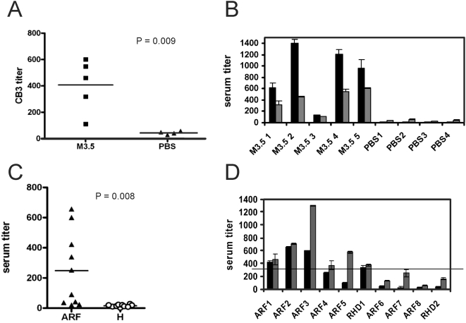
Acute rheumatic fever (ARF) and rheumatic heart disease are serious autoimmune sequelae to infections with Streptococcus pyogenes. Streptococcal M-proteins have been implicated in ARF pathogenesis. Their interaction with collagen type IV (CIV) is a triggering step that induces generation of collagen-specific auto-antibodies. Electron microscopy of the protein complex between M-protein type 3 (M3-protein) and CIV identified two prominent binding sites of which one is situated in the CB3-region of CIV. In a radioactive binding assay, M3-protein expressing S. pyogenes and S. gordonii bound the CB3-fragment. Detailed analysis of the interactions by surface plasmon resonance measurements and site directed mutagenesis revealed high affinity interactions with dissociation constants in the nanomolar range that depend on the recently described collagen binding motif of streptococcal M-proteins. Because of its role in the induction of disease-related collagen autoimmunity the motif is referred to as "peptide associated with rheumatic fever" (PARF). Both, sera of mice immunized with M3-protein as well as sera from patients with ARF contained anti-CB3 auto-antibodies, indicating their contribution to ARF pathogenesis. The identification of the CB3-region as a binding partner for PARF directs the further approaches to understand the unusual autoimmune pathogenesis of PARF-dependent ARF and forms a molecular basis for a diagnostic test that detects rheumatogenic streptococci.


Conflict of interest statement

Competing Interests: The authors have declared that no competing interests exist.

Figures

Figure 1
Figure 1. Electron microscopy of M3-CIV complexes.
Micrographies (left panel) and corresponding cartoons (right panel) show complexes that consist of M3-protein and CIV. Block arrows highlight M3-protein (depicted in gray in the cartoons), both free and bound to CIV (depicted in black in the cartoons). Arrow heads point out the 7S regions of CIV, while line arrows points out the globular heads of the CIV molecules. The bars represent 100 nm in the representative overviev (A) and 50 nm in the panel of selected complexes (B).
Figure 2
Figure 2. CB3 binding by M3-protein.
(A) Interaction with 125I-CB3 was measured in a pull-down assay with, M3-GST (black bar), or M18-GST (grey bar) protein coupled to glutathion sepharose beads. A control sample with glutathion separose alone (−) was included (white bar). (B) Binding of 125I-CIV (black bars) or 125I-CB3 (grey bars) to S. gordonii that heterologously expressed M3 protein on its surface (SGO M3) and that of the wild type control (SGO) was expressed as a percentage of total radioactivity input. Error bars in A and B represent the standard deviation of the triplicate measurements. (C and D) Surface plasmon resonance measurements of the interaction between M3-protein and immobilized CIV (C) or CB3 (D), respectively. Injection of M3-protein at different concentrations (a: 50 µg/ml, b: 25 µg/ml, c: 12.5 µg/ml, d: 6.25 µg/ml) started at t = 0 s and stopped at t = 120 s. The response is expressed in response units (RU).
Figure 3
Figure 3. Binding of PARF mutants to CB3.
(A) Dot blot experiment with 125I-CB3 as the soluble ligand. Dots 1–9 are the immobilized mutants of M protein FOG (for description see table in B); dot 10 is recombinant wild type protein. Recombinant M3- (+) and M18-proteins (−) were used as positive and negative control, respectively. The table in B allows to compare the experiment with previous data on full length collagen IV.
Figure 4
Figure 4. CIV and CB3 specific serum responses.
(A) Serum Ig response for CB3 in M3.5 immunized mice. Reactivity was determined for sera from PBS-immunized (PBS) or M3-protein-immunized (M3.5) mice. The anti-CIV (black bars) and anti-CB3 titers (grey bars) measured in individual mice immunized with M3.5 (M3.5 #) or PBS (PBS #) are compared in (B). (C) Anti-CB3 titer of individual patient sera. Titers are shown for each individual serum collected from ARF/RHD patients (triangles), or healthy individuals (circles). The significance of the differences measured in A and C was examined using the t-test. (D) The six sera of patients with CB3 autoimmunity (black bars) showed also reactivity against the collagen binding part of M3-protein (M3.5) (grey bars). The horizontal line indicates the cut-off titer for M3.5 positive sera. Error bars represent the standard deviation of the triplicate measurements in B an D.

Similar articles

Cited by

References

    1. Cunningham MW. Pathogenesis of group A streptococcal infections. Clin Microbiol Rev. 2000;13:470–511. - PMC - PubMed
    1. WHO. Rheumatic fever and rheumatic heart disease. World Health Organization. 2004:923 923. - PubMed
    1. Carapetis JR, McDonald M, Wilson NJ. Acute rheumatic fever. Lancet. 2005;366:155–168. - PubMed
    1. Hudson BG, Tryggvason K, Sundaramoorthy M, Neilson EG. Alport's syndrome, Goodpasture's syndrome, and type IV collagen. N Engl J Med. 2003;348:2543–2556. - PubMed
    1. Dinkla K, Nitsche-Schmitz DP, Barroso V, Reissmann S, Johansson HM, et al. Identification of a streptococcal octapeptide motif involved in acute rheumatic fever. J Biol Chem. 2007;282:18686–18693. - PubMed

Publication types