

Mammalian cell display for antibody engineering
- PMID: 19252852
- PMCID: PMC3475965
- DOI: 10.1007/978-1-59745-554-1_18
Mammalian cell display for antibody engineering
Abstract
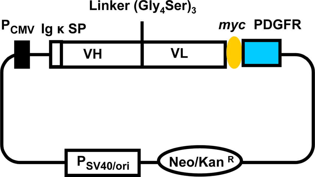
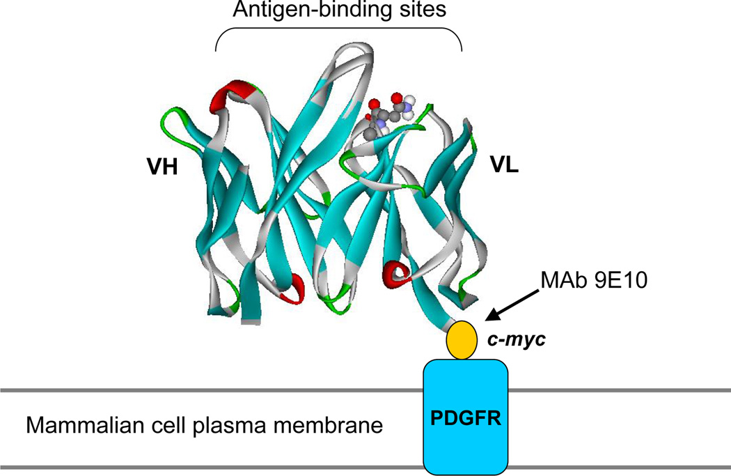
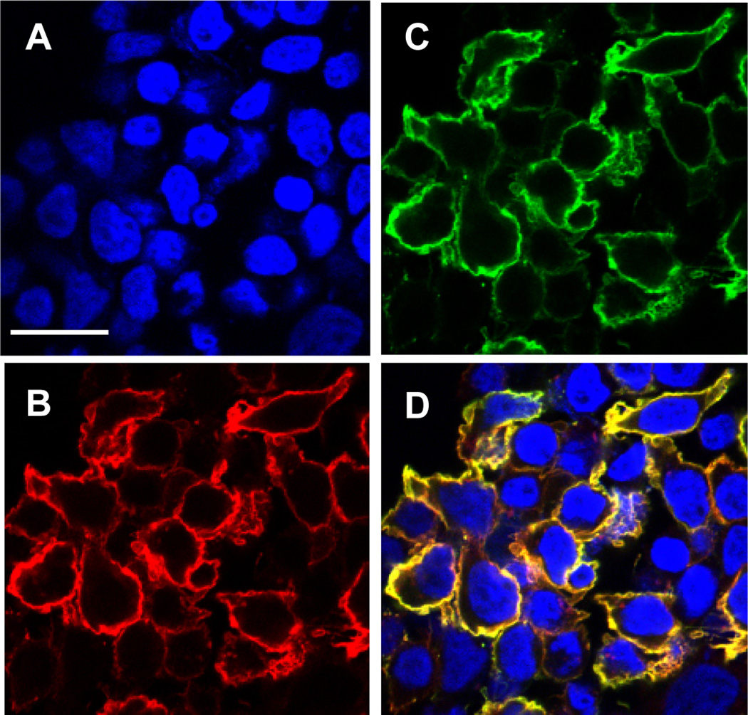
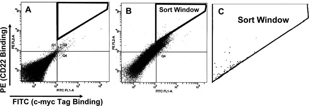
Antibody engineering has generally been carried out by displaying mouse or human antibodies or antibody fragments on the surface of microorganisms (phage, bacteria, and yeast). We have shown that mammalian cells can be used to display single-chain antibody fragments (scFvs) for affinity maturation. Using mammalian cell display one can isolate and engineer scFvs, Fabs, or whole IgGs for increased affinity and other specific biological functions. Here, we describe a mammalian cell display strategy to isolate high-affinity scFvs specific for CD22. Our strategy uses flow cytometry and human embryonic kidney 293T (HEK-293T) cells that are widely used for transient protein expression. Flow cytometry enhances the screen's sensitivity thereby allowing us to isolate high-affinity antibodies.
Figures
References
-
- Adams GP, Weiner LM. Monoclonal antibody therapy of cancer. Nat. Biotechnol. 2005;23:1147–1157. - PubMed
-
- Smith GP. Filamentous fusion phage: novel expression vectors that display cloned antigens on the virion surface. Science. 1985;228:1315–1317. - PubMed
-
- Winter G, Griffiths AD, Hawkins RE, Hoogenboom HR. Making antibodies by phage display technology. Annu. Rev. Immunol. 1994;12:433–455. - PubMed
-
- Boder ET, Wittrup KD. Yeast surface display for screening combinatorial polypeptide libraries. Nat. Biotechnol. 1997;15:553–557. - PubMed
Publication types
MeSH terms
Substances
Grants and funding
LinkOut - more resources
Full Text Sources
Other Literature Sources
Miscellaneous
