. 2009 May;52(5):891-900.
doi: 10.1007/s00125-009-1294-y. Epub 2009 Feb 28.

Potential role of TBC1D4 in enhanced post-exercise insulin action in human skeletal muscle

Affiliations

Potential role of TBC1D4 in enhanced post-exercise insulin action in human skeletal muscle

J T Treebak et al. Diabetologia. 2009 May.

Abstract

Aims/hypothesis: TBC1 domain family, member 4 (TBC1D4; also known as AS160) is a cellular signalling intermediate to glucose transport regulated by insulin-dependent and -independent mechanisms. Skeletal muscle insulin sensitivity is increased after acute exercise by an unknown mechanism that does not involve modulation at proximal insulin signalling intermediates. We hypothesised that signalling through TBC1D4 is involved in this effect of exercise as it is a common signalling element for insulin and exercise.

Methods: Insulin-regulated glucose metabolism was evaluated in 12 healthy moderately trained young men 4 h after one-legged exercise at basal and during a euglycaemic-hyperinsulinaemic clamp. Vastus lateralis biopsies were taken before and immediately after the clamp.

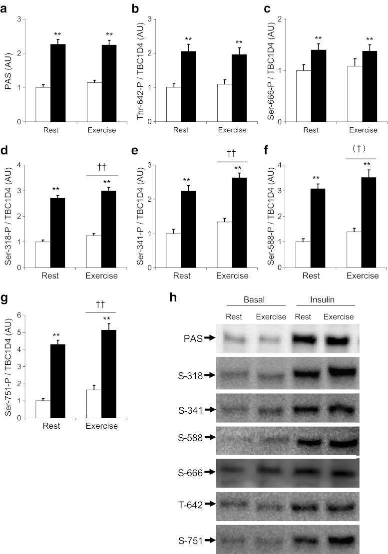
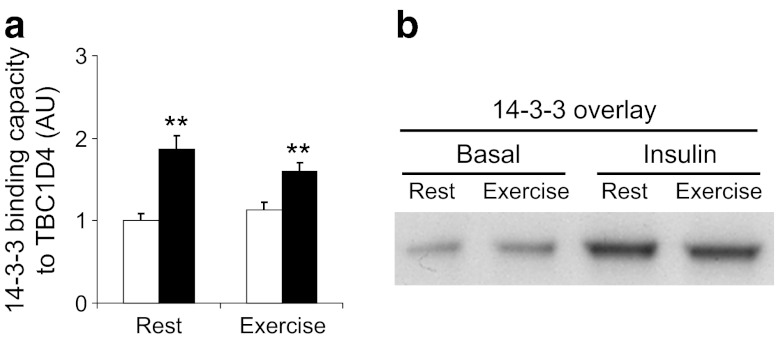
Results: Insulin stimulation increased glucose uptake in both legs, with greater effects (approximately 80%, p < 0.01) in the previously exercised leg. TBC1D4 phosphorylation, assessed using the phospho-AKT (protein kinase B)substrate antibody and phospho- and site-specific antibodies targeting six phosphorylation sites on TBC1D4, increased at similar degrees to insulin stimulation in the previously exercised and rested legs (p < 0.01). However, TBC1D4 phosphorylation on Ser-318, Ser-341, Ser-588 and Ser-751 was higher in the previously exercised leg, both in the absence and in the presence of insulin (p < 0.01; Ser-588, p = 0.09; observed power = 0.39). 14-3-3 binding capacity for TBC1D4 increased equally (p < 0.01) in both legs during insulin stimulation.

Conclusion/interpretation: We provide evidence for site-specific phosphorylation of TBC1D4 in human skeletal muscle in response to physiological hyperinsulinaemia. The data support the idea that TBC1D4 is a nexus for insulin- and exercise-responsive signals that may mediate increased insulin action after exercise.


Figures

Fig. 1
Fig. 1
TBC1D4 phosphorylation in the post-exercise state. TBC1D4 phosphorylation was evaluated in vastus lateralis muscle samples obtained in the basal state 4 h after termination of a one-legged knee extensor exercise bout (white bars) and at the end of a euglycaemic–hyperinsulinaemic clamp (black bars) using the PAS antibody and phospho-specific antibodies targeting six of the known phosphorylation sites on TBC1D4. Values, in arbitrary units (AU), are for (a) PAS, (b) Thr-642, (c) Ser-666, (d) Ser-318, (e) Ser-341, (f) Ser-588 and (g) Ser-751. **p < 0.01 for insulin effect, n = 12; ††p < 0.01 for prior exercise effect, n = 12; (†)p = 0.09 borderline significance, observed power = 0.39, n = 12. h Representative blots using TBC1D4 phospho-specific antibodies. All antibodies gave a single band running at 160 kDa. Ser-666 was measured after immunoprecipitation of TBC1D4; all other blots were direct blots performed using vastus lateralis muscle lysates
Fig. 2
Fig. 2
Binding capacity of 14–3–3 to TBC1D4 increased with insulin as shown in (a). Following immunoprecipitation of TBC1D4, 14–3–3 binding capacity to TBC1D4 was evaluated by far-western overlay (b) using digoxigenin-labelled 14–3–3 protein. **p < 0.01 for insulin effect, n = 12
Fig. 3
Fig. 3
Verification of antibodies specificity. a TBC1D1 and TBC1D4 were immunoprecipitated from a human vastus lateralis muscle sample and the immunoprecipitates (IP) (150 μg) loaded together with the pre-IPs (lysate cleared with beads; 30 μg) and the post-IPs (supernatant fraction; 30 μg). Following SDS-PAGE proteins were transferred to PVDF membranes, which were then probed with TBC1D4 antibody. b Same samples as above (a), but the membranes were incubated with TBC1D1 antibody. c Membranes (a) were re-incubated with a mixture of TBC1D1 and TBC1D4 antibodies and developed to show the separation between TBC1D1 and TBC1D4. d Western blot analyses were performed using an insulin-stimulated human vastus lateralis muscle sample loaded in three wells. The membrane was cut through the middle lane. The two membranes were incubated with different antibodies as indicated. The different pieces of membrane were put back together and developed. TBC1D1 and TBC1D4 ran at different molecular masses, whereas the signal from the PAS antibody aligned with TBC1D4, but not with TBC1D1. e TBC1D4 was immunoprecipitated from the same sample used above (d), and pre-IP (60 μg), post-IP (60 μg) and IP (120 μg) were subjected to SDS-PAGE and transferred to PVDF membranes. These were then incubated with antibodies as indicated. All bands in the IP lane ran with the same, or slightly higher, molecular mass as the pre-IP sample. IB, immunoblot
Fig. 4
Fig. 4
a mRNA expression of human TBC1D1 and TBC1D4 splice variants in skeletal muscle and subcutaneous adipose tissue. Relative amounts of the different gene products were calculated and compared. Adipose tissue (white bars) exclusively expressed the short (-S) version of TBC1D4 and TBC1D1. Expression of TBC1D1 was approximately threefold higher than that of TBC1D4 in adipose tissue. Skeletal muscle (black bars) expressed equal amounts of TBC1D4-long (-L) and TBC1D1, but only very small amounts of TBC1D4-S, which may be contamination from inter-myocellular fat cells. b TBC1D4 Western blot showing that human skeletal muscle (M) and subcutaneous adipose tissue (A) differentially produce TBC1D4-L and TBC1D4-S splice variant proteins. **p < 0.01 vs TBC1D4-S within tissue, (n = 8); ††p < 0.01 for within gene, n = 8. IB, immunoblot

References

    1. Richter EA, Garetto LP, Goodman MN, Ruderman NB. Muscle glucose metabolism following exercise in the rat: increased sensitivity to insulin. J Clin Invest. 1982;69:785–793. doi: 10.1172/JCI110517. - DOI - PMC - PubMed
    1. Bogardus C, Thuillez P, Ravussin E, Vasquez B, Narimiga M, Azhar S. Effect of muscle glycogen depletion on in vivo insulin action in man. J Clin Invest. 1983;72:1605–1610. doi: 10.1172/JCI111119. - DOI - PMC - PubMed
    1. Garetto LP, Richter EA, Goodman MN, Ruderman NB. Enhanced muscle glucose metabolism after exercise in the rat: the two phases. Am J Physiol Endocrinol Metab. 1984;246:E471–E475. - PubMed
    1. Richter EA, Garetto LP, Goodman MN, Ruderman NB. Enhanced muscle glucose metabolism after exercise: modulation by local factors. Am J Physiol Endocrinol Metab. 1984;246:E476–E482. - PubMed
    1. Mikines KJ, Sonne B, Farrell PA, Tronier B, Galbo H. Effect of physical exercise on sensitivity and responsiveness to insulin in humans. Am J Physiol Endocrinol Metab. 1988;254:E248–E259. - PubMed

Publication types

MeSH terms