Review
. 2009 May;29(3):247-56.
doi: 10.1007/s10875-009-9278-8. Epub 2009 Feb 28.

Cytokine-producing dendritic cells in the pathogenesis of inflammatory skin diseases

Affiliations
Review

Cytokine-producing dendritic cells in the pathogenesis of inflammatory skin diseases

Leanne M Johnson-Huang et al. J Clin Immunol. 2009 May.

Abstract

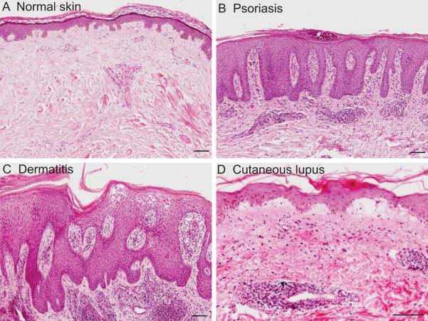
Introduction: Inflammatory skin diseases can be examined from many viewpoints. In this review, we consider three distinct cutaneous inflammatory diseases from the point of view of their major lesional dendritic cell (DC) subpopulations. The DC populations considered are Langerhans cells, myeloid DCs, and plasmacytoid DCs (pDCs), with specific attention to the presence and role of the inflammatory counterparts of these cells. From such a "dendritic cell-centric" focus, psoriasis, atopic dermatitis (AD), and cutaneous lupus erythematosus (CLE) are explored.

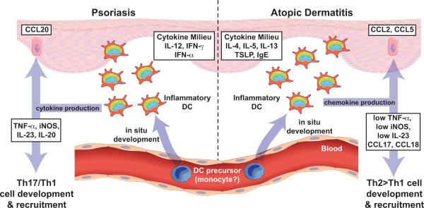
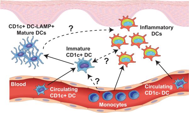
Discussion: In psoriasis, there is a specific population of myeloid "inflammatory" DCs that appears to play an important pathogenic role, while pDCs have been recently implicated in the initiation of psoriatic lesions. In AD, Langerhans cells may be important during initiation, while "inflammatory dendritic epidermal cells" (IDECs) appear to be abundant in lesional epidermis and dermis and contribute to maintenance of AD. These IDECs may actually be analogous to the myeloid inflammatory DCs found in the epidermal and dermal compartments of the skin in psoriasis, although they express distinct surface markers and induce different T cell polarities as a result of different cytokine milieu in which they develop. CLE has been recently characterized as a type I IFN-mediated disease, and pDCs are integral to the pathogenesis of this disease.

Conclusion: Thus, these DC subpopulations and their products will be reviewed in the context of these three cutaneous diseases to provide clinico-pathophysiological correlations between the lesional DCs, their products, and the skin diseases.


Figures

Figure 1
Figure 1. Normal skin, psoriasis, atopic dermatitis, and cutaneous lupus tissue sections by haematoxylin and eosin staining
(A) Normal skin can be divided into the epidermis, the upper outer layer of keratinocyte cells with a “basket weave” stratum corneum, and the dermis, the pale pink layer beneath the epidermis. (B) Psoriasis is characterized by a greatly thickened epidermis (acanthosis) with elongation of the rete ridges that protrude down into the dermis. There is retention of keratinocyte nuclei in the outer layers (parakeratosis). Collections of neutrophils can also be seen in the epidermis and stratum corneum. In the dermis, there is elongation and dilatation of the dermal blood vessels that protrude up between the rete ridges. These dermal blood vessels and surrounding upper dermal spaces are filled with predominantly mononuclear leukocytes (including DCs and T cells) and neutrophils. (C) Dermatitis may produce epidermal edema, which breaks apart the epidermal connections (spongiosis). The epidermis becomes acanthotic with scratching, and there is a variable dermal infiltrate consisting of T cells, DCs, macrophages, eosinophils and mast cells. (D) Acute cutaneous lupus skin lesions demonstrate characteristic involvement of the dermo-epidermal junction that separates the epidermis and dermis. Lymphocytes line up and can obscure this region in a “lichenoid” histological pattern. In an acute presentation, there is edema associated with this region as shown here. There is a mixed dermal infiltrate associated with this process including lymphocytes and DCs, which may be patchy or surround vessels and appendages.
Figure 2
Figure 2. The development and function of myeloid inflammatory DCs in psoriasis and AD
As a result of cytokine production in the cutaneous microenvironment, DC precursors differentiate into inflammatory DCs in situ. Depending on the environmental cues, inflammatory DCs produce a distinct set of cytokines. Psoriatic DCs produce TNF-α, nitric oxide, IL-20 and IL-23 and are involved in the development and maintenance of a Th1/Th17 polarized response. In AD, inflammatory DCs neither produce much TNF or IL-23, nor have an iNOS signature, but instead produce chemokines, CCL17 and CCL18, that are involved in ongoing Th2>Th1 cell recruitment.
Figure 3
Figure 3. The putative precursors of inflammatory DCs
It is unclear which cells give rise to inflammatory DCs. It is possible that inflammatory DCs are derived from monocytes infiltrating the skin, or they could differentiate from resident or infiltrating CD1c+ DCs.

References

    1. Zaba LC, Krueger JG, Lowes MA. Resident and “inflammatory” dendritic cells in human skin. J Invest Dermatol. 2009;129:302–8. - PMC - PubMed
    1. Klechevsky E, Morita R, Liu M, Cao Y, Coquery S, Thompson-Snipes L, Briere F, Chaussabel D, Zurawski G, Palucka AK, Reiter Y, Banchereau J, Ueno H. Functional specializations of human epidermal Langerhans cells and CD14+ dermal dendritic cells. Immunity. 2008;29:497–510. - PMC - PubMed
    1. Merad M, Ginhoux F, Collin M. Origin, homeostasis and function of Langerhans cells and other langerin-expressing dendritic cells. Nat Rev Immunol. 2008;8:935–47. - PubMed
    1. Zaba LC, Fuentes-Duculan J, Steinman RM, Krueger JG, Lowes MA. Normal human dermis contains distinct populations of CD11c+BDCA-1+ dendritic cells and CD163+FXIIIA+ macrophages. J Clin Invest. 2007;117:2517–25. - PMC - PubMed
    1. Ito T, Amakawa R, Inaba M, Hori T, Ota M, Nakamura K, Takebayashi M, Miyaji M, Yoshimura T, Inaba K, Fukuhara S. Plasmacytoid dendritic cells regulate Th cell responses through OX40 ligand and type I IFNs. J Immunol. 2004;172:4253–9. - PubMed

Publication types

MeSH terms