. 2009 Mar 23;10(5):862-8.
doi: 10.1002/cbic.200800805.

Molecular assembly of an aptamer-drug conjugate for targeted drug delivery to tumor cells

Affiliations

Molecular assembly of an aptamer-drug conjugate for targeted drug delivery to tumor cells

Yu-Fen Huang et al. Chembiochem. .

Abstract

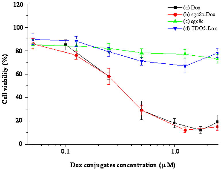
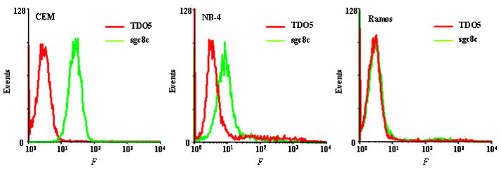
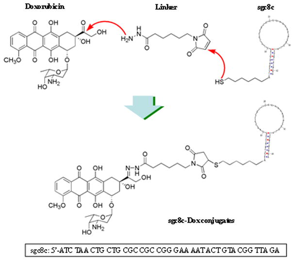
The conjugation of antitumor drugs to targeting reagents such as antibodies is a promising method that can increase the efficacy of chemotherapy and reduce the overall toxicity of the drugs. In this study, we covalently link the antitumor agent doxorubicin (Dox) to the DNA aptamer sgc8c, which was selected by the cell-SELEX method. In doing so, we expected that this sgc8c-Dox conjugate would specifically kill the target CCRF-CEM (T-cell acute lymphoblastic leukemia, T-cell ALL) cells, but with minimal toxicity towards nontarget cells. The results demonstrated that the sgc8c-Dox conjugate possesses many of the properties of the sgc8c aptamer, including high binding affinity (K(d)=2.0+/-0.2 nM) and the capability to be efficiently internalized by target cells. Moreover, due to the specific conjugation method, the acid-labile linkage connecting the sgc8c-Dox conjugate can be cleaved inside the acidic endosomal environment. Cell viability tests demonstrate that the sgc8c-Dox conjugates not only possess potency similar to unconjugated Dox, but also have the required molecular specificity that is lacking in most current targeted drug delivery strategies. Furthermore, we found that nonspecific uptake of membrane-permeable Dox to nontarget cell lines could also be inhibited by linking the drug with the aptamer; thus, the conjugates are selective for cells that express higher amounts of target proteins. Compared to the less effective Dox-immunoconjugates, these sgc8c-Dox conjugates make targeted chemotherapy more feasible with drugs having various potencies. When combined with the large number of recently created DNA aptamers that specifically target a wide variety of cancer cells, this drug-aptoconjugation method will have broad implications for targeted drug delivery.


Figures

Figure 1
Figure 1
Binding assay of sgc8c-Dox conjugates to CCRF-CEM cells. A) Flow cytometry assay for the binding of sgc8c-Dox conjugates with CCRF-CEM cells. The red and green curves represent the fluorescence from fluorescein-sgc8c incubated with pure cells and cells labeled with sgc8c-Dox (200 nM), respectively. B) Flow cytometry to determine the binding affinity of sgc8c-Dox conjugates to CCRF-CEM cells. The fluorescence is derived from the second stain of cells by fluorescein-labeled sgc8c. Inset is the plot of mean fluorescence (a. u.) versus sgc8c-Dox concentration (nM) in log scale.
Figure 2
Figure 2
Cytotoxicity assays of (a) sgc8c-Dox conjugates, (b) free Dox, (c) free sgc8c, and (d) TDO5-Dox conjugates with CCRF-CEM cell line. The cells (2 × 10⁴ cells/well) were incubated with Dox or aptamer-Dox conjugates (0 to 2.5 μM) in culture medium without FBS at 37 °C, 5% CO2 for 2 h. After drug treatment, cells were subsequently grown in fresh medium (10% FBS) for 48 h. The cytotoxicity was then measured by MTT assay.
Figure 3
Figure 3
Confocal images display the distribution of sgc8c-Dox conjugates inside CCRF-CEM cells at different time points: A) 30 min, B) 1 h, and C) 2 h. The cells (106) were incubated with sgc8c-Dox (0.5 μM) and transferrin-Alexa633 (60 nM) in culture medium without FBS at 37 °C, 5% CO2 for 2 h. From left to right, the fluorescence images were monitored for sgc8c-Dox, transferrin-alexa633, overlay of these two channels, and bright field channel, respectively. Transferrin-Alexa633 will both bind to the surface and internalize to the endosomal compartment of CCRF-CEM cells.
Figure 4
Figure 4
Quantitative analysis of A) Dox and B) sgc8c-Dox inside CCRF-CEM cells. The cells (105 cells) were incubated with Dox or aptamer-Dox conjugates in culture medium without FBS at 37 °C, 5% CO2 for 2 h. A) Dashed and solid lines represent the fluorescence signal from Dox before and after trypsin treatment, respectively. Trypsin was applied to remove the membrane-bound Dox. B) Fluorescence signal from sgc8c-Dox inside cells determined by flow cytometry. All the signals have been subtracted by the fluorescence intensity from TDO5-Dox nonspecifically uptaken by CCRF-CEM cells.
Figure 5
Figure 5
Cytotoxicity assays of free Dox, sgc8c-Dox, and TDO5-Dox conjugates with two additional cell lines: A) NB-4 and B) Ramos cells. All the conditions are the same as those in Figure 2.
Figure 6
Figure 6
Flow cytometry assay for the binding of biotin-labeled TDO5 and sgc8c with three different cell lines: CCRF-CEM, NB-4, and Ramos cells. Cells (105) were incubated with biotin-labeled TDO5 and sgc8c at 37 °C for 20 min in 100 μL culture medium without FBS. After washing twice, cells were mixed with streptavidin-(R-phycoerythrin) (20 min on ice), and the fluorescence was determined by flow cytometry.
Scheme 1
Scheme 1
Conjugation of the drug doxorubicin (Dox) to aptamer sgc8c for targeted delivery to cancer cells.

References

    1. Carter PJ, Senter PD. Cancer J. 2008;14:154–169. - PubMed
    1. Chari RVJ. Acc Chem Res. 2008;41:98–107. - PubMed
    1. Chu TC, Marks JW, Lavery LA, Faulkner S, Rosenblum MG, Ellington AD, Levy M. Cancer Res. 2006;66:5989–5992. - PubMed
    1. Bagalkot V, Farokhzad OC, Langer R, Jon S. Angew Chem Int Ed. 2006;45:8149–8152. - PubMed
    1. Farokhzad OC, Karp JM, Langer R. Expert Opin Drug Deliv. 2006;3:311–324. - PubMed

Publication types

MeSH terms

LinkOut - more resources