. 2009 Mar 2:4:8.
doi: 10.1186/1749-8104-4-8.

Cytoplasmic polyadenylation and cytoplasmic polyadenylation element-dependent mRNA regulation are involved in Xenopus retinal axon development

Affiliations

Cytoplasmic polyadenylation and cytoplasmic polyadenylation element-dependent mRNA regulation are involved in Xenopus retinal axon development

Andrew C Lin et al. Neural Dev. .

Abstract

Background: Translation in axons is required for growth cone chemotropic responses to many guidance cues. Although locally synthesized proteins are beginning to be identified, how specific mRNAs are selected for translation remains unclear. Control of poly(A) tail length by cytoplasmic polyadenylation element (CPE) binding protein 1 (CPEB1) is a conserved mechanism for mRNA-specific translational regulation that could be involved in regulating translation in axons.

Results: We show that cytoplasmic polyadenylation is required in Xenopus retinal ganglion cell (RGC) growth cones for translation-dependent, but not translation-independent, chemotropic responses in vitro, and that inhibition of CPE binding through dominant-negative interference severely reduces axon outgrowth in vivo. CPEB1 mRNA transcripts are present at low levels in RGCs but, surprisingly, CPEB1 protein was not detected in eye or brain tissue, and CPEB1 loss-of-function does not affect chemotropic responses or pathfinding in vivo. UV cross-linking experiments suggest that CPE-binding proteins other than CPEB1 in the retina regulate retinal axon development.

Conclusion: These results indicate that cytoplasmic polyadenylation and CPE-mediated translational regulation are involved in retinal axon development, but that CPEB1 may not be the key regulator of polyadenylation in the developing retina.


Figures
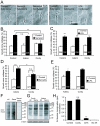
Figure 1
Figure 1
Cytoplasmic polyadenylation is required for growth cone collapse. (A) Typical examples of collapsed and non-collapsed growth cones. (B) Cordycepin (200 μM; 3'deoxyadenosine; Cordy), but not adenosine (Adeno), blocks Sema3A-induced growth cone collapse. (C) Cordycepin does not affect lysophosphatidic acid (LPA)-induced growth cone collapse. (D, E) Similar results are obtained as in (B, C) if axons are severed from their cell bodies. (F, G) Anti-puromycin western blots on stage 35/36 retinal cultures incubated for 15 minutes with 2 μM puromycin and 50 μM LnLL (F) or 0.5 μM puromycin (G), ± 2 μg/ml Sema3A-Fc, ± 40 μM anisomycin (aniso), ± 200 μM cordycepin or adenosine. Cordycepin slightly reduces, but does not abolish, Sema3A-induced translation (G); see text for details. The two separate sections in (F) are from the same blot with the same film exposure and contrast settings. (H) Cordycepin does not block basal translation in A6 fibroblasts, while cycloheximide (CHX) does. Numbers in bars indicate number of growth cones counted. *p < 0.05, **p < 0.01, ***p < 0.001. Error bars represent standard error of the mean.
Figure 2
Figure 2
CPEB1 mRNA is expressed in the Xenopus embryonic retina. (A) RT-PCR shows that CPEB1 is expressed in stage 35/36 and stage 41 eyes and that the level of expression is higher at stage 41. (B, C) Wholemount in situ hybridization on stage 35/36 (B) and stage 41 (C) embryos shows that CPEB1 is weakly expressed in the eyes, as well as the nasal placode and brain, and in a segmented pattern in the dorsal spinal cord. The sense probe gives no signal. AS, antisense; np, nasal placode; S, sense; sc, spinal cord. (D) Section of stage 41 eye before (D) and after (D') laser capture microdissection of the retinal ganglion cell layer. Abbreviations: inl, inner nuclear layer; le, lens; onl, outer nuclear layer; rgc, retinal ganglion cell layer; rpe, retinal pigment epithelium. (E) RT-PCR on RNA from the retinal ganglion cell layer isolated by laser capture microdissection shows that CPEB1 is expressed in retinal ganglion cells. (F) CPEB2–4 are also expressed in embryonic eyes and brains by RT-PCR.
Figure 3
Figure 3
CPEB1 loss-of-function does not cause obvious retinal axon guidance defects. (A) Anti-green fluorescent protein (GFP) western blot on lysates of stage 24 embryos injected with 1 ng CPEB1-RBM-GFP mRNA with or without CPEB1 morpholino (MO) at the two-cell stage. CPEB1 MO effectively knocks down expression of CPEB1-GFP. (B) DiI filling of the retinal projection of stage 41 Xenopus embryos injected with CPEB1 MO at the two-cell stage shows no obvious defect in in vivo axon pathfinding. Uninj, uninjected. (C-G) Blastomere injection (C-D) and electroporation (EP) (E-G) of CPEB1 MO does not affect Sema3A-induced growth cone collapse. Blastomere injection (C) and electroporation (E, F) of carboxyfluorescein-tagged CPEB1 MO labels cell bodies (E) and growth cones (C, G) containing the MO with green fluorescence. (H) Beta-tubulin staining of wholemount mouse retinas shows no obvious defect in intraretinal guidance in embryonic day (E)18 CPEB1 knockout mice; retinal axons converge normally on the optic disc. (I, J) DiI filling of the retinal projection at E18–19 reveals no obvious defect in retinal axon guidance at the optic chiasm (F) or in the optic tract (G). Abbreviations: ch, optic chiasm; dLGN, dorsal lateral geniculate nucleus; sc, superior colliculus. Error bars represent standard error of the mean.
Figure 4
Figure 4
Xenopus embryonic retinas contain non-CPEB1 cytoplasmic polyadenylation element (CPE)-binding proteins. (A) Western blots using three different antibodies do not detect CPEB1 protein in stage 45 eyes, stage 45 heads, stage 41 eyes, or stage 35/6 eyes. However, they do detect CPEB1 in stage VI oocytes. pc, polyclonal antibody against amino terminus of Xenopus CPEB1; 2B7, monoclonal antibody against human CPEB1; pept, polyclonal antibody against amino-terminal peptide of Xenopus CPEB1 (see Materials and methods). (B) All three antibodies used detect recombinant CPEB1 in stage 24 embryos injected with CPEB1-RBM-GFP mRNA (Figure 3A). MO, morpholino. (C) UV cross-linking with 32P-labeled CPE-containing probe suggests the presence of non-CPEB1 CPE-binding proteins. Stage 41 eye extracts were incubated with 32P-UTP labeled probe consisting of the 3' end of Xenopus cyclin B1 with CPE motifs intact (lane 1) or mutated (lane 2), and resolved by SDS-PAGE. Two bands are labeled with 32P (upper panel; indicated by arrows) but not detected by anti-CPEB1 western blot (lower panel). Proteins binding only to the mutated probe (lane 2) may recognize sequences masked by the CPE-binding proteins in the intact CPE-containing probe. Cross-linked samples were also subject to immunoprecipitation (IP) by control IgG (lane 3) or anti-CPEB1 antibody (lanes 4–5; pc, the polyclonal antibody labeled 'pc' in (A)). Anti-CPEB1 did not immunoprecipitate any cross-linked 32P-labeled CPE probe (lane 4). The bands at 70 kDa and 50 kDa on the western blot are most likely non-specific, as they do not correspond to any 32P signal. WB, western blot.
Figure 5
Figure 5
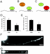
The CPEB1 mutant (S174A, S180A) (CPEB1-AA) disrupts neurite outgrowth in vitro. (A-C) Model for how CPEB1-AA competitively interferes with the function of endogenous cytoplasmic polyadenylation element (CPE)-binding proteins. Normally, unidentified CPE-binding proteins regulate the translation or localization of CPE-containing mRNAs (A). The CEPB1-RNA binding mutant (C529A, C539A) (CPEB1-RBM), being unable to bind to CPE-containing RNAs, does not affect this situation and thus serves as a negative control (B). However, CPEB1-AA binds to the CPE, displacing native CPE-binding proteins, thus preventing CPE-mediated mRNA regulation (C). (D) Fewer retinal cells transfected with CPEB1-AA-GFP (AA) have neurites compared to cells transfected with CPEB1-RBM-GFP (RBM). (E) AA-expressing dissociated retinal neurons have shorter neurites than those expressing RBM. Neurite length was quantified by digitally tracing the longest neurite of each transfected neuron (see Materials and methods for details). (F, G) Dissociated retinal neurons transfected with RBM (F), or AA (G). Note the presence of CPEB1-GFP in the neurites. *p < 0.05; ***p < 0.001. Scale bars: 15 μm. Error bars represent standard error of the mean.
Figure 6
Figure 6
The CPEB1 mutant (S174A, S180A) (CPEB1-AA) disrupts axon outgrowth in vivo. (A) Retinal ganglion cells (RGCs) transfected with CPEB1-RBM-GFP (RBM) send axons to the optic tectum correctly. ch, optic chiasm. (B) Embryos where RGCs are transfected with CPEB1-AA-GFP (AA) do not have GFP-positive axons in the brain. (C) Schematic of bidirectional plasmid encoding both GAP-red fluorescent protein (RFP) and CPEB1-green fluorescent protein (GFP). CMV, cytomegalovirus; mPGK, mouse phosphoglycerate kinase. (D-G) Electroporation of bidirectional plasmids causes co-expression of GAP-RFP and CPEB1-GFP. Dashed boxes in (D) and (F) are shown at higher resolution in (E) and (G), respectively. (H) Diagram of optic pathway in horizontal sections. Dashed boxes indicate the part of the pathway shown in (I), (J), and (K). (I) RFP/RBM- and RFP/AA-transfected axons in the optic nerve head. These axons contain CPEB1-GFP (arrows). (J) Very rarely, an RFP/AA-transfected axon extends just beyond the optic chiasm. Dashed lines indicate expected path of retinal axons. (K) RFP/RBM-transfected, but not RFP/AA-transfected, axons reach and branch in the tectum (arrowhead). The RFP/RBM image is a composite of two adjacent sections. (L) Number of RFP/RBM- and RFP/AA-transfected embryos with detectable RFP-positive axons reaching each intermediate target, analyzed in sections. (M-N) RFP/RBM-transfected embryos have bright RFP-positive axons in the optic pathway projecting correctly to the tectum, while RFP/AA-transfected embryos do not. Scale bars: 65 μm (A, B, M, N); 10 μm (D, F, J, K); 5 μm (I). Blue, DAPI; green, CPEB1-GFP; red, GAP-RFP. Dashed lines indicate inner and outer plexiform layers and solid lines indicate the apical and basal limits of the retina. Abbreviations: INL, inner nuclear layer; ONL, outer nuclear layer.
Figure 7
Figure 7
The CPEB1 mutant (S174A, S180A) (CPEB1-AA) reduces, but does not abolish, retinal ganglion cell (RGC) differentiation. (A) Cells transfected with CPEB1-GFP classified into the six retinal cell types according to layering and morphology; no differences were observed. (B) Examples of cells in the RGC layer transfected with CPEB1-RBM-GFP (RBM) or CPEB1-AA-GFP (AA) that are positive for Isl-1 staining. Blue, DAPI; green, GFP; red, Isl-1. In each image, the upper and lower dotted lines indicate the outer and inner plexiform layers, respectively. The upper and lower solid lines indicate the apical and basal limits of the retina, respectively. Numbers in or above bars indicate number of cells counted. (C) Fewer cells in the RGC layer transfected with AA express Isl-1 compared to RGCs transfected with RBM. However, approximately half of AA-expressing cells still express Isl-1. Scale bars 5 μm. Abbreviations: A, amacrine cell; BP, bipolar cell; GCL, ganglion cell layer; H, horizontal cell; INL, inner nuclear layer; M, Müller cell; ONL, outernuclear layer; PR, photoreceptor; RPE, retinal pigment epithelium.

Similar articles

Cited by

References

    1. Dickson BJ. Molecular mechanisms of axon guidance. Science. 2002;298:1959–1964. doi: 10.1126/science.1072165. - DOI - PubMed
    1. Campbell DS, Holt CE. Chemotropic responses of retinal growth cones mediated by rapid local protein synthesis and degradation. Neuron. 2001;32:1013–1026. doi: 10.1016/S0896-6273(01)00551-7. - DOI - PubMed
    1. Brunet I, Weinl C, Piper M, Trembleau A, Volovitch M, Harris W, Prochiantz A, Holt C. The transcription factor Engrailed-2 guides retinal axons. Nature. 2005;438:94–98. doi: 10.1038/nature04110. - DOI - PMC - PubMed
    1. Piper M, Anderson R, Dwivedy A, Weinl C, van Horck F, Leung KM, Cogill E, Holt C. Signaling mechanisms underlying Slit2-induced collapse of Xenopus retinal growth cones. Neuron. 2006;49:215–228. doi: 10.1016/j.neuron.2005.12.008. - DOI - PMC - PubMed
    1. Lin AC, Holt CE. Function and regulation of local axonal translation. Curr Opin Neurobiol. 2008;18:60–68. doi: 10.1016/j.conb.2008.05.004. - DOI - PMC - PubMed

Publication types

MeSH terms