Review
. 2009 Mar 2:6:7.
doi: 10.1186/1743-0003-6-7.

Transcranial magnetic stimulation, synaptic plasticity and network oscillations

Affiliations
Review

Transcranial magnetic stimulation, synaptic plasticity and network oscillations

Patricio T Huerta et al. J Neuroeng Rehabil. .

Abstract

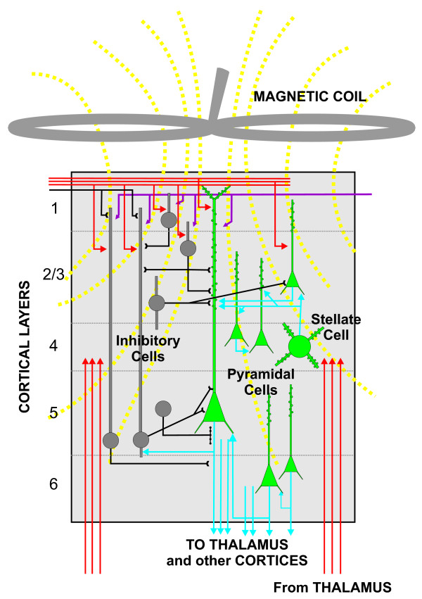
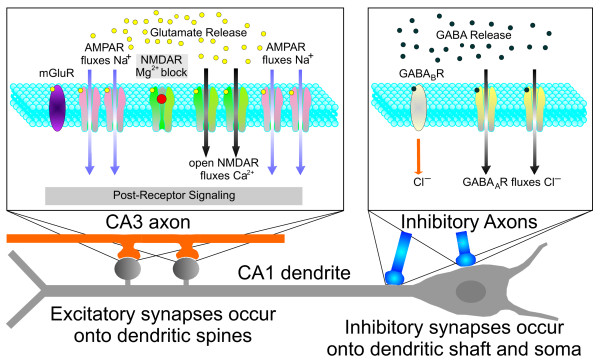
Transcranial magnetic stimulation (TMS) has quickly progressed from a technical curiosity to a bona-fide tool for neurological research. The impetus has been due to the promising results obtained when using TMS to uncover neural processes in normal human subjects, as well as in the treatment of intractable neurological conditions, such as stroke, chronic depression and epilepsy. The basic principle of TMS is that most neuronal axons that fall within the volume of magnetic stimulation become electrically excited, trigger action potentials and release neurotransmitter into the postsynaptic neurons. What happens afterwards remains elusive, especially in the case of repeated stimulation. Here we discuss the likelihood that certain TMS protocols produce long-term changes in cortical synapses akin to long-term potentiation and long-term depression of synaptic transmission. Beyond the synaptic effects, TMS might have consequences on other neuronal processes, such as genetic and protein regulation, and circuit-level patterns, such as network oscillations. Furthermore, TMS might have non-neuronal effects, such as changes in blood flow, which are still poorly understood.


Figures

Figure 1
Figure 1
Schematic representation of the human cerebral cortex. The magnetic coil, represented as a figure-of-eight device, is placed on top of the cerebral cortex and pulses a magnetic field that induces electrical currents across the six layers of the cerebral cortex (indicated by numbers at left). The excitatory cells (green with blue axons) and the inhibitory cells (gray with black axons) have the potential to be activated at the level of their axons, which contain the highest density of ion channels. The incoming axons from other cortical areas and the thalamus (indicated in red) are also activated. The end result of the magnetic pulse is the synaptic activation of a chain of neurons, which generate feed-forward and feedback loops of excitation and inhibition.
Figure 2
Figure 2
Schematic representation of the glutamatergic and GABAergic receptors in a CA1 pyramidal neuron. The left box represents a CA3-CA1 synapse. The CA3 axon (orange) releases glutamate from the presynaptic terminals. The postsynaptic CA1 neuron expresses three types of glutamatergic receptors: metabotropic receptor (mGluR), alpha-amino-3-hydroxy-5-methyl-4-isoxazolepropionic acid receptor (AMPAR), and N-methyl-D-aspartate receptor (NMDAR). The AMPARs are represented in their active state, as they allow Na+ to enter onto the dendritic spine. The NMDARs are represented both in the closed state (leftmost NMDAR, with the Mg2+ block seen as a red ball in the mouth of the receptor) and in the open state, when the NMDARs allow Ca2+ to enter onto the spine (notice the absence of the Mg2+ block). The right box represents a synapse between an inhibitory interneuron and the CA1 cell. The interneuron releases γ-aminobutyric acid (GABA) onto the CA1 pyramidal neuron, which expresses GABAA receptors (yellow) and GABAB receptors (gray), leading to inhibition of the target cell. The GABAA receptors are represented in the open state when they allow Cl- to enter onto the CA1 dendrite.

References

    1. Barker AT, Jalinous R, Freeston IL. Non-invasive magnetic stimulation of human motor cortex. Lancet. 1985;1:1106–1107. doi: 10.1016/S0140-6736(85)92413-4. - DOI - PubMed
    1. Hallett M. Transcranial magnetic stimulation: a primer. Neuron. 2007;55:187–199. doi: 10.1016/j.neuron.2007.06.026. - DOI - PubMed
    1. Pascual-Leone A, Davey N, Rothwell J, Wassermann EM, Puri BK. Handbook of Transcranial Magnetic Stimulation. London: Hodder Arnold; 2002.
    1. Walsh V, Pascual-Leone A. Transcranial Magnetic Stimulation: A Neurochronometrics of Mind. Cambridge: The MIT Press; 2005.
    1. Wassermann E, Epstein C, Ziemann U. Oxford Handbook of Transcranial Stimulation. Oxford: Oxford University Press; 2008.

Publication types

MeSH terms

LinkOut - more resources