. 2009 Mar 4;96(5):1733-50.
doi: 10.1016/j.bpj.2008.11.050.

Quantitative modeling and analysis of the transforming growth factor beta signaling pathway

Affiliations

Quantitative modeling and analysis of the transforming growth factor beta signaling pathway

Seung-Wook Chung et al. Biophys J. .

Abstract

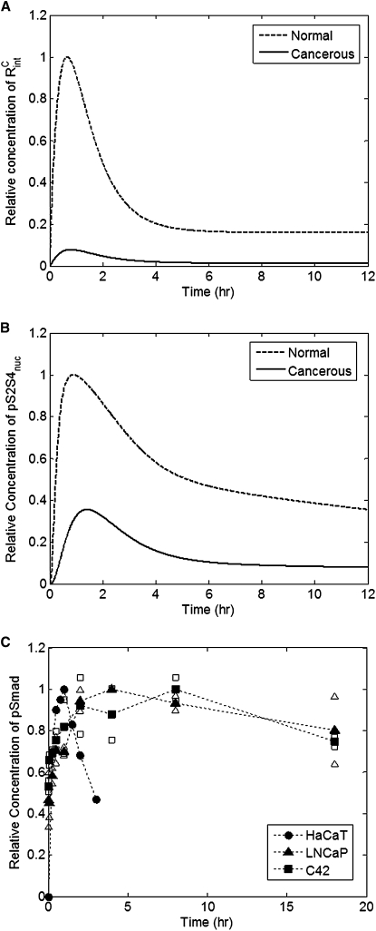
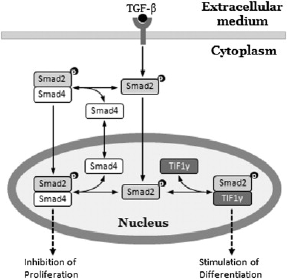
Transforming growth factor beta (TGF-beta) signaling, which regulates multiple cellular processes including proliferation, apoptosis, and differentiation, plays an important but incompletely understood role in normal and cancerous tissues. For instance, although TGF-beta functions as a tumor suppressor in the premalignant stages of tumorigenesis, paradoxically, it also seems to act as a tumor promoter in advanced cancer leading to metastasis. The mechanisms by which TGF-beta elicits such diverse responses during cancer progression are still not entirely clear. As a first step toward understanding TGF-beta signaling quantitatively, we have developed a comprehensive, dynamic model of the canonical TGF-beta pathway via Smad transcription factors. By describing how an extracellular signal of the TGF-beta ligand is sensed by receptors and transmitted into the nucleus through intracellular Smad proteins, the model provides quantitative insight into how TGF-beta-induced responses are modulated and regulated. Subsequent model analysis shows that mechanisms associated with Smad activation by ligand-activated receptor, nuclear complex formation among Smad proteins, and inactivation of ligand-activated Smad (e.g., degradation, dephosphorylation) may be critical for regulating TGF-beta-targeted functional responses. The model was also used to predict dynamic characteristics of the Smad-mediated pathway in abnormal cells, from which we generated four testable hypotheses regarding potential mechanisms by which TGF-beta's tumor-suppressive roles may appear to morph into tumor-promotion during cancer progression.


Figures

Figure 1
Figure 1
Schematic representation of the pathway components in the integrated model. Numbers in the cartoon refer to the chemical reaction indices in Table 1.
Figure 2
Figure 2
Model fit to experimental data: (A) total phosphorylated Smad2 in the nucleus (20), (B) total phosphorylated Smad2 in the cytoplasm (30), (C) total nuclear Smad2 (30), (D) total cytoplasmic Smad2 (30), and (E) total nuclear Smad4 (30) in response to TGF-β stimulation. Note: to simulate the effects of cycloheximide in (B–E), all rates of protein synthesis (k12syn, k13syn, k14syn, and k15syn) were set to zero.
Figure 3
Figure 3
Model validation: (A) total cellular pSamd2 (43); (B) ratio of cellular pSmad2 to total Smad2 in response to the step input of TGF-β (41); (C) ratio of cellular pSmad2 to total Smad2 in response to the pulse input of TGF-β (41); (D) total cytoplasmic Smad4 (30). To simulate the effects of cycloheximide in (D), all rates of protein synthesis (k12syn, k13syn, k14syn, and k15syn) were set to zero.
Figure 4
Figure 4
Model parameter sensitivities for select parameters with the greatest influence on phosphorylated Smad2-Smad4 complex in the nucleus. Parameters with maximum normalized sensitivity coefficients exceeding 0.5 in absolute value at any point in time are shown.
Figure 5
Figure 5
The effect of blocking translocation of monomeric pSmad2 (dashed, k17imp = 0) or heteromeric pSmad2 (dash dot, k7imp = 0).
Figure 6
Figure 6
The effect of variations in the rate of (A) Smad2 phosphorylation, (B) nuclear pSmad2-Smad4 association, (C) pSmad2 degradation, (D) nuclear pSmad2 dephosphorylation, (E) and nuclear import of pSmad2 on the dynamics of nuclear pSmad2-Smad4 complex. Each indicated parameter value was increased (dashed) or decreased (dash dot) 10-fold.
Figure 7
Figure 7
The effect of different concentrations of TGF-β on the dynamic responses of (A) internalized activated receptor complex and (B) activated Smad2-Smad4 complex in the nucleus under normal conditions, and (C) internalized activated receptor complex and (D) activated Smad2-Smad4 complex in the nucleus under cancerous conditions with 10-fold reduction in initial levels and protein synthesis rate constants of both Type I and Type II receptors. (E) Maximum responses of activated nuclear Smad2-Smad4 complex to different doses of TGF-β (0.02, 0.1, 0.2, 1, 2, 10, and 20 pM) in normal cells (open circles) and cancerous cells (solid triangles), respectively.
Figure 8
Figure 8
In silico mutation results. Responses of (A) internalized activated receptor complex and (B) nuclear pSmad-Smad4 complex to 10-fold reduction in initial levels and protein synthesis rate constants of both Type I and Type II receptors. (C) Temporal profiles of phosphorylated Smad2 in HaCaT cells (circles, from Lo and Massague (43)), LNCaP cells (triangles, our experiments), and C4-2 cells (squares, our experiments) in response to 200 pM (for A) or 400 pM (for B and C) of TGF-β. The open triangles and squares represent the maximum and minimum of the data at each time point; the solid squares and triangles denote the corresponding average values. All data points were normalized with respect to the maximum intensity value of pSmad2 of each profile.
Figure 9
Figure 9
Model predictions for (A) nuclear Smad4 and (B) nuclear pSmad2-Smad4 complex on TGF-β stimulation (80 pM) under cancerous conditions; 10-fold reduction in the initial levels and the protein synthesis rate constants of both Type I and Type II receptors, and 10-fold increase in rates of degradation of either Smad4 (A, squares; B, diamonds) or pSmad2 (B, triangles) or both (B, squares).
Figure 10
Figure 10
Alternative TGF-β-induced responses determined by nuclear pSmad2-binding partners. Whereas Smad4 forms transcriptional complexes with receptor-phosphorylated Smad2/3 and mediates antiproliferative responses, TIF1γ specifically recognizes receptor-activated Smad2/3 and mediates differentiation of hematopoietic stem/progenitor cells. (Adapted from He et al. (54)).

Similar articles

Cited by

References

    1. Massague J. TGF-β signal transduction. Annu. Rev. Biochem. 1998;67:753–791. - PubMed
    1. Massague J., Gomis R.R. The logic of TGFβ signaling. FEBS Lett. 2006;580:2811–2820. - PubMed
    1. Shi Y., Massague J. Mechanisms of TGF-β signaling from cell membrane to the nucleus. Cell. 2003;113:685–700. - PubMed
    1. Massague J. How cells read TGF-β signals. Nat. Rev. Mol. Cell Biol. 2000;1:169–178. - PubMed
    1. Itoh S., Itoh F., Goumans M.J., Ten Dijke P. Signaling of transforming growth factor-β family members through Smad proteins. Eur. J. Biochem. 2000;267:6954–6967. - PubMed

Publication types