

Cross-correlated TIRF/AFM reveals asymmetric distribution of force-generating heads along self-assembled, "synthetic" myosin filaments
- PMID: 19254555
- PMCID: PMC2717282
- DOI: 10.1016/j.bpj.2008.11.032
Cross-correlated TIRF/AFM reveals asymmetric distribution of force-generating heads along self-assembled, "synthetic" myosin filaments
Abstract
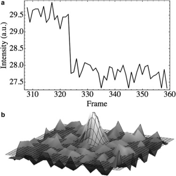
Myosin-II's rod-like tail drives filament assembly with a head arrangement that is often considered to be a symmetric bipole that generates equal and opposite contractile forces on actin. Self-assembled myosin filaments are shown here to be asymmetric in physiological buffer based on cross-correlated images from both atomic force microscopy and total internal reflection fluorescence. Quantitative cross-correlation of these orthogonal methods produces structural information unavailable to either method alone in showing that fluorescence intensity along the filament length is proportional to height. This implies that myosin heads form a shell around the filament axis, consistent with F-actin binding. A motor density of approximately 50-100 heads/micrometer is further estimated but with an average of 32% more motors on one half of any given filament compared to the other, regardless of length. A purely entropic pyramidal lattice model is developed and mapped onto the Dyck paths problem that qualitatively captures this lack of length dependence and the distribution of filament asymmetries. Such strongly asymmetric bipoles are likely to produce an unbalanced contractile force in cells and in actin-myosin gels and thereby contribute to motility as well as cytoskeletal tension.
Figures


Similar articles
-
Myosin thick filaments from adult rabbit skeletal muscles.Biochim Biophys Acta. 1999 Nov 16;1472(3):413-30. doi: 10.1016/s0304-4165(99)00136-1. Biochim Biophys Acta. 1999. PMID: 10564756
-
Multiple- and single-molecule analysis of the actomyosin motor by nanometer-piconewton manipulation with a microneedle: unitary steps and forces.Biophys J. 1996 Jan;70(1):383-400. doi: 10.1016/S0006-3495(96)79582-6. Biophys J. 1996. PMID: 8770215 Free PMC article.
-
Atomic force microscopy of thermally treated myosin filaments.J Agric Food Chem. 2005 Jun 1;53(11):4589-92. doi: 10.1021/jf0500381. J Agric Food Chem. 2005. PMID: 15913330
-
Packing of myosin molecules in muscle thick filaments.Cell Biol Int. 2000;24(6):327-33. doi: 10.1006/cbir.1999.0514. Cell Biol Int. 2000. PMID: 10860567 Review.
-
Cargo recognition and cargo-mediated regulation of unconventional myosins.Acc Chem Res. 2014 Oct 21;47(10):3061-70. doi: 10.1021/ar500216z. Epub 2014 Sep 17. Acc Chem Res. 2014. PMID: 25230296 Review.
Cited by
-
The power of correlative microscopy: multi-modal, multi-scale, multi-dimensional.Curr Opin Struct Biol. 2011 Oct;21(5):686-93. doi: 10.1016/j.sbi.2011.06.010. Epub 2011 Jul 21. Curr Opin Struct Biol. 2011. PMID: 21782417 Free PMC article.
-
Progress in the Correlative Atomic Force Microscopy and Optical Microscopy.Sensors (Basel). 2017 Apr 24;17(4):938. doi: 10.3390/s17040938. Sensors (Basel). 2017. PMID: 28441775 Free PMC article. Review.
-
Controlling the Electromagnetic Field Confinement with Metamaterials.Sci Rep. 2016 Nov 25;6:37739. doi: 10.1038/srep37739. Sci Rep. 2016. PMID: 27886230 Free PMC article.
-
Construction of a Three-Color Prism-Based TIRF Microscope to Study the Interactions and Dynamics of Macromolecules.Biology (Basel). 2021 Jun 23;10(7):571. doi: 10.3390/biology10070571. Biology (Basel). 2021. PMID: 34201434 Free PMC article.
-
Imaging with total internal reflection fluorescence microscopy for the cell biologist.J Cell Sci. 2010 Nov 1;123(Pt 21):3621-8. doi: 10.1242/jcs.056218. J Cell Sci. 2010. PMID: 20971701 Free PMC article. Review.
References
-
- Conti M.A., Adelstein R.S. Nonmuscle myosin II moves in new directions. J. Cell Sci. 2008;121:11–18. - PubMed
-
- Ip K., Sobieszek A., Solomon D., Jiao Y., Par P.D. Physical integrity of smooth muscle myosin filaments is enhanced by phosphorylation of the regulatory myosin light chain. Cell. Physiol. Biochem. 2007;20:649–658. - PubMed
-
- Decker B., Kellermayer M.S.Z. Periodically arranged interactions within the myosin filament backbone revealed by mechanical unzipping. J. Mol. Biol. 2008;377:307–310. - PubMed
-
- Davis J.S. Assembly processes in vertebrate skeletal thick filament formation. Annu. Rev. Biophys. Biophys. Chem. 1988;17:217–239. - PubMed
-
- Sellers J.R., Kachar B. Polarity and velocity of sliding filaments—control of direction by actin and of speed by myosin. Science. 1990;249:406–408. - PubMed
Publication types
MeSH terms
Substances
LinkOut - more resources
Full Text Sources
Miscellaneous
