. 2009 Mar;9(3):217-27.
doi: 10.1016/j.cmet.2009.01.010.

Interaction of the hereditary hemochromatosis protein HFE with transferrin receptor 2 is required for transferrin-induced hepcidin expression

Affiliations

Interaction of the hereditary hemochromatosis protein HFE with transferrin receptor 2 is required for transferrin-induced hepcidin expression

Junwei Gao et al. Cell Metab. 2009 Mar.

Abstract

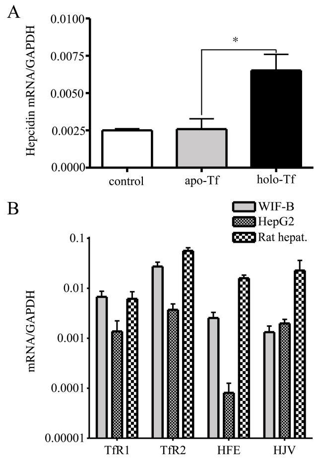
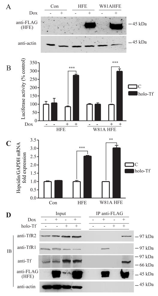
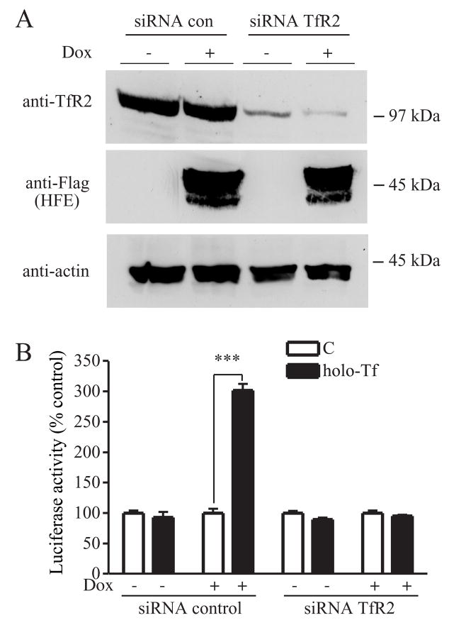
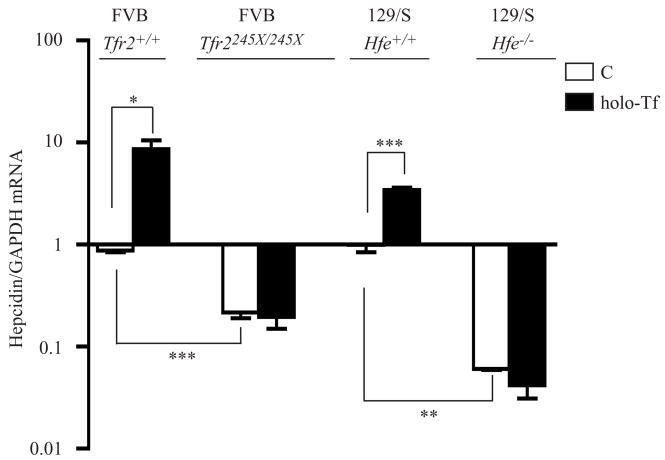
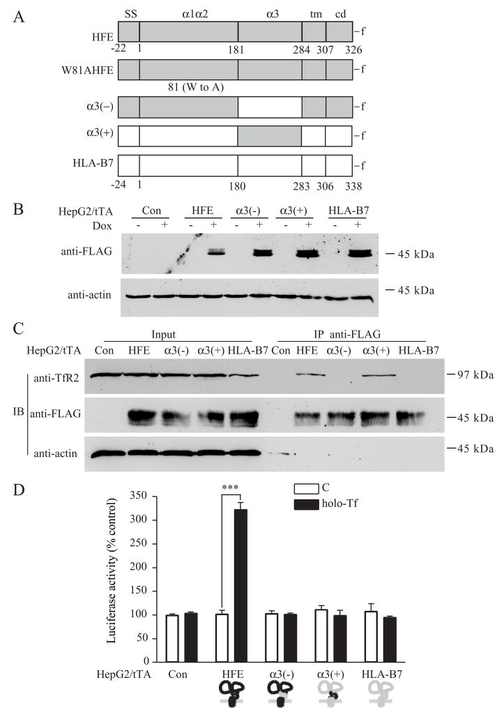
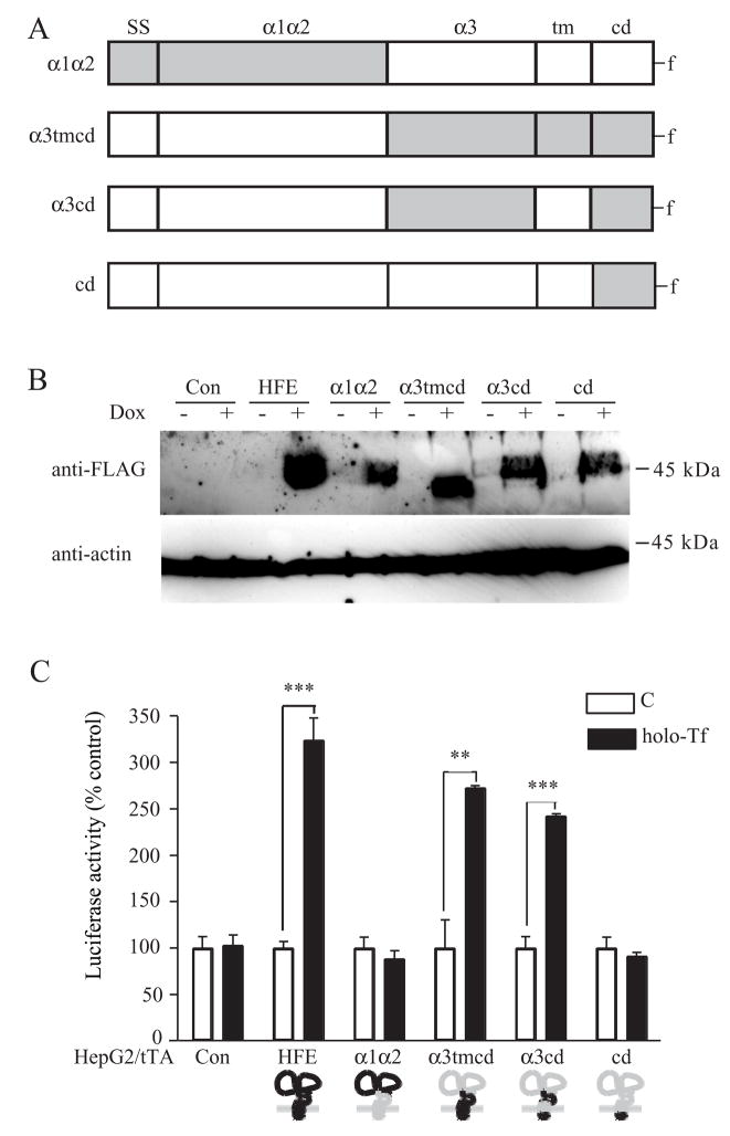
The mechanisms that allow the body to sense iron levels in order to maintain iron homeostasis are unknown. Patients with the most common form of hereditary iron overload have mutations in the hereditary hemochromatosis protein HFE. They have lower levels of hepcidin than unaffected individuals. Hepcidin, a hepatic peptide hormone, negatively regulates iron efflux from the intestines into the blood. We report two hepatic cell lines, WIF-B cells and HepG2 cells transfected with HFE, where hepcidin expression responded to iron-loaded transferrin. The response was abolished when endogenous transferrin receptor 2 (TfR2) was suppressed or in primary hepatocytes lacking either functional TfR2 or HFE. Furthermore, transferrin-treated HepG2 cells transfected with HFE chimeras containing only the alpha3 and cytoplasmic domains could upregulate hepcidin expression. Since the HFE alpha3 domain interacts with TfR2, these results supported our finding that TfR2/HFE complex is required for transcriptional regulation of hepcidin by holo-Tf.


Figures

Figure 1
Figure 1. Hepcidin expression in WIF-B cells responds to holo-Tf
A. Holo-Tf induces hepcidin expression in WIF-B cells. WIF-B cells were left untreated or incubated with 25 μM human apo-Tf or holo-Tf for 24 hours, prior to RNA isolation. The hepcidin mRNA level was measured by qRT-PCR and normalized to GAPDH. B. Comparison of HFE, TfR1, TfR2 and HJV mRNA levels in WIF-B cells, HepG2 cells, and rat hepatocytes. Levels of TfR1, TfR2, HFE and HJV mRNA, iron related genes upstream of hepcidin, were measured by qRT-PCR. All samples were run in triplicate in three independent experiments. Data are shown as average ± S.D. P-values < 0.05 are indicated by *.
Figure 2
Figure 2. Tf-induced dissociation of HFE from TfR1 results in the association of HFE with TfR2 and stimulates hepcidin promoter activity
A. Inducible expression of wild-type or W81A mutant HFE in HepG2/tTA HFE and HepG2/tTA W81AHFE cells. Lysates (25 μg) of HepG2/tTA HFE or HepG2/tTA W8AHFE cells, uninduced (−dox) or induced (+dox) to express HFE or W81AHFE, were detected with anti-FLAG antibody. B. HFE is required for Tf to induce hepcidin promoter activity independently of interaction with TfR1. Cells were cotransfected with pLuc-link-HAMP and pCMV-β-gal, treated without (C) or with holo-Tf and analyzed as described in Experimental Procedures. Results are expressed as average ± S.D. of three independent experiments performed in triplicate. C. Hepcidin mRNA expression in HepG2/tTA (Con) HepG2/tTA HFE (HFE) and HepG2/tTA W81AHFE (W81AHFE) cells were treated without (C) or with holo-Tf as described in Experimental Procedures. The expression of hepcidin was measured by qRT-PCR and normalized to GAPDH. The fold of increase in hepcidin mRNA in the dox-induced group was obtained by normalization to uninduced controls. All samples were run in triplicate in three independent experiments. Data are shown as average ± S.D. D. Tf releases HFE from TfR1 to bind to TfR2 in HepG2 cells. Cell lysates (200 μg) from HepG2/tTA HFE treated with or without 25 μM holo-Tf were immunoprecipitated with rabbit anti-FLAG antibodies, and Sepharose-4B/Protein A. Proteins were detected on immunoblots using mouse anti- TfR2, TfR1, FLAG, and actin and goat anti-Tf. The input lanes correspond to 1/5 of the material used for immunoprecipitation. These results were repeated once with similar results. P-values < 0.001 are indicated by *** and < 0.01 by **.
Figure 3
Figure 3. Knockdown of TfR2 blocks Tf-mediated induction of hepcidin expression in HepG2/tTA HFE cells
A. Knockdown of endogenous TfR2 in HepG2/tTA HFE cells using specific siRNA to TfR2. Cells were transfected with control siRNA or TfR2 siRNA and co-transfected with pLuc-link-HAMP and pCMV-β-gal as described in Experimental Procedures. To observe the efficiency of knockdown of TfR2, cell lysates (50 μg) from TfR2 siRNA or control siRNA transfected HepG2/tTA HFE cells were analyzed by immunoblot for TfR2, HFE and actin expression. These results were representative of one out of three experiments. Human TfR2 siRNA significantly decreased endogenous TfR2 protein level in both HepG2/tTA control cells and HepG2/tTA HFE cells. B. Hepcidin promoter activity assay in HepG2/tTA HFE cells after knockdown of TfR2 and treatment without (C) or with holo-Tf. Luciferase and β-galactosidase activity from cell lysates were measured as described in Experimental Procedures. Relative luciferase activity was obtained by normalization to β-galactosidase activity and the fold increase in luciferase activity of each group was obtained by normalization to the control group without holo-Tf. Results are expressed as average ± S.D. of three independent experiments performed in triplicate. P-values < 0.001 are indicated by ***.
Figure 4
Figure 4. TfR2 and HFE are required for Tf-mediated induction of hepcidin expression in mouse primary hepatocytes
Mouse primary hepatocytes were treated without (C) or with holo-Tf and qRT-PCR was performed on isolated RNA as described in Experimental Procedures. Average relative level of hepcidin was normalized to GAPDH. Results are expressed as average ± S.D. of two or four independent experiments performed in triplicate. P-values < 0.05, < 0.01 and < 0.001 are indicated by *, ** and ***, respectively.
Figure 5
Figure 5. The α3 domain is required but insufficient for HFE-mediated Tf induction of hepcidin expression in HepG2 cells
A. Schematic representation of the HFE mutant and chimeras including HFE, W81AHFE, α3(−),α3(+) and HLA-B7. HFE is shown in gray, HLA-B7 is shown in white. Numbers under diagrams for each full-length protein designate the first amino acid in the signal sequence (SS), α1α2 domain (α1α2), α3 domain (α3), transmembrane domain (tm), and cytoplasmic domain (cd). FLAG epitope tag (-f) was added to the C terminus of each protein. HFE-HLA-B7 chimeras were constructed by replacing the appropriate segments between the parent proteins. The first coding methionine in all proteins is labeled as number 1. We refer to individual recombinant proteins used throughout the study by acronyms to left of the construct illustration. B. Induction of HFE chimeras by dox. Lysates (25 μg) of HepG2/tTA HFE, HepG2/tTA α3(−), HepG2/tTA α3(+) and HepG2/tTA HLA-B7 cells, uninduced (−dox) or induced (+dox) to express wtHFE, α3(−),α3(+) or HLA-B7 were detected using mouse anti-FLAG antibody. C. The α3 domain of HFE is critical for interaction with TfR2 in HepG2/tTA cells. Cells were treated without (C) or with 25 μM holo-Tf (holo-Tf) as described in Experimental Procedures. Cells lysates (200 μg) were immunoprecipitated with rabbit anti-FLAG antibody and the blots were probed with mouse antibodies to TfR2, FLAG and actin. The input lanes correspond to 1/5 of the material used for immunoprecipitation. These results were repeated once with similar results. D. The α3 domain of HFE is required but insufficient for HFE-mediated Tf induction of hepcidin expression in HepG2 cells. Cells were cotransfected with pLuc-link-HAMP and pCMV-β-gal plasmids, treated without (C) or with 25 μM holo-Tf (holo-Tf), and analyzed as described in Experimental Procedures. Results are expressed as average ± S.D. of three independent experiments performed in triplicate. P-values < 0.001 are indicated by ***.
Figure 6
Figure 6. The α3 and cytoplasmic domains of HFE mediate the Tf induction of hepcidin expression in HepG2 cells
A. Schematic representation of the HFE chimeras including α1α2, α3tmcd, α3cd and cd constructs. HFE is shown in gray, HLA-B7 is shown in white. HFE chimeras were constructed by replacing the appropriate segments between the parent proteins. Individual recombinant proteins used throughout the study are referred to by the acronyms to left of the construct cartoon. B. Generation of HFE-HLA-B7 chimeras. HFE chimeras including stable cells lines HepG2/tTA α1α2, HepG2/tTA α3tmcd, HepG2/tTA α3cd and HepG2/tTA cd were generated as described in Experimental Procedures. Lysates (25 μg) of cells, uninduced (−dox) or induced (+dox) were used to screen the positive clones by immunoblot using anti-FLAG antibody. C. The α3 and cytoplasmic domain of HFE is required for HFE-mediated Tf induction of hepcidin expression in HepG2 cells. All HepG2/tTA control and HFE chimeras cells were cotransfected with pLuc-link-HAMP and pCMV-β-gal, treated, and analyzed as described in Experimental Procedures. Results are expressed as average ± S.D. of three independent experiments performed in triplicate. P-values < 0.001 are indicated by *** and < 0.01 by **.

Comment in

References

    1. Ahmad KA, Ahmann JR, Migas MC, Waheed A, Britton RS, Bacon BR, Sly WS, Fleming RE. Decreased liver hepcidin expression in the Hfe knockout mouse. Blood Cells Mol Dis. 2002;29:361–366. - PubMed
    1. Ajioka RS, Kushner JP. Hereditary hemochromatosis. Semin Hematol. 2002;39:235–241. - PubMed
    1. Bennett MJ, Lebron JA, Bjorkman PJ. Crystal structure of the hereditary haemochromatosis protein HFE complexed with transferrin receptor. Nature. 2000;403:46–53. - PubMed
    1. Bondi A, Valentino P, Daraio F, Porporato P, Gramaglia E, Carturan S, Gottardi E, Camaschella C, Roetto A. Hepatic expression of hemochromatosis genes in two mouse strains after phlebotomy and iron overload. Haematologica. 2005;90:1161–1167. - PubMed
    1. Bothwell TH, MacPhail AP. Hereditary hemochromatosis: etiologic, pathologic, and clinical aspects. Semin Hematol. 1998;35:55–71. - PubMed

Publication types

MeSH terms