. 2009 Mar;9(3):277-86.
doi: 10.1016/j.cmet.2009.01.006.

Adipocyte CREB promotes insulin resistance in obesity

Affiliations

Adipocyte CREB promotes insulin resistance in obesity

Ling Qi et al. Cell Metab. 2009 Mar.

Abstract

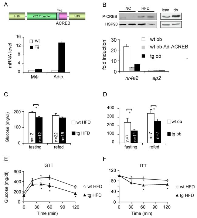
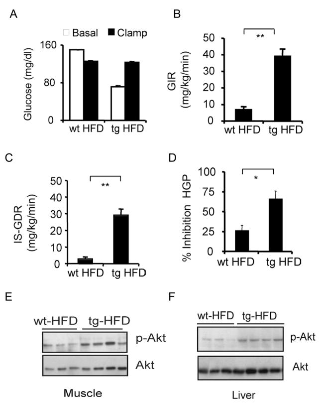
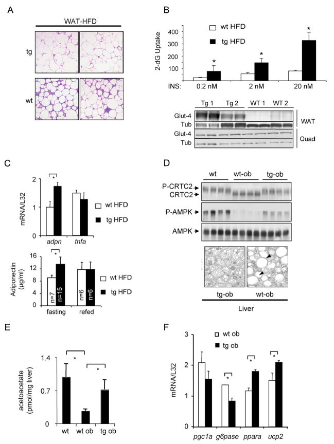
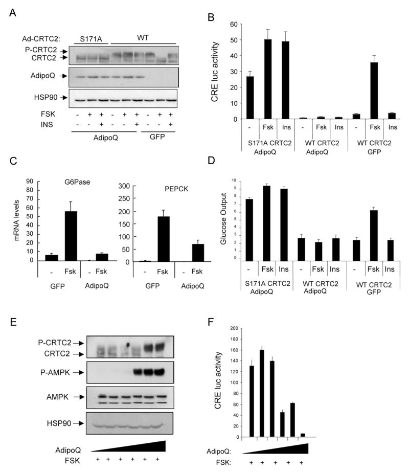
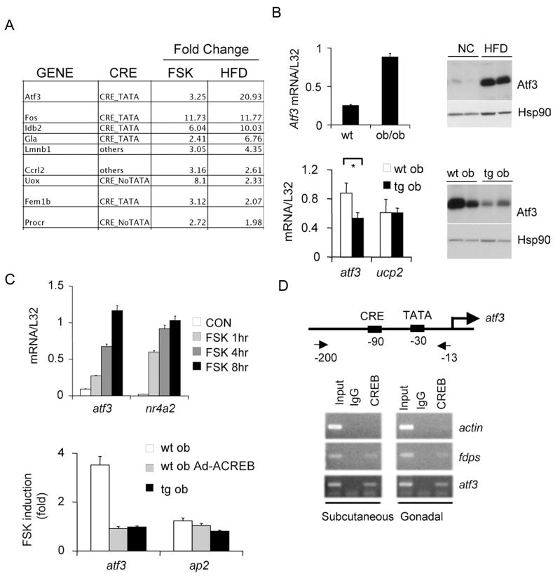
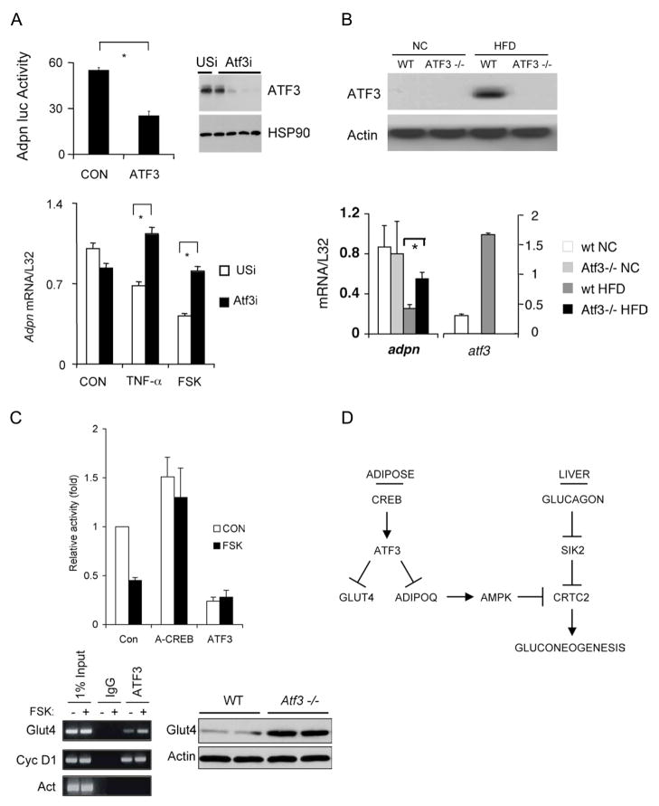
Increases in adiposity trigger metabolic and inflammatory changes that interfere with insulin action in peripheral tissues, culminating in beta cell failure and overt diabetes. We found that the cAMP Response Element Binding protein (CREB) is activated in adipose cells under obese conditions, where it promotes insulin resistance by triggering expression of the transcriptional repressor ATF3 and thereby downregulating expression of the adipokine hormone adiponectin as well as the insulin-sensitive glucose transporter 4 (GLUT4). Transgenic mice expressing a dominant-negative CREB transgene in adipocytes displayed increased whole-body insulin sensitivity in the contexts of diet-induced and genetic obesity, and they were protected from the development of hepatic steatosis and adipose tissue inflammation. These results indicate that adipocyte CREB provides an early signal in the progression to type 2 diabetes.


Figures

Figure 1
Figure 1
Mice deficient in adipocyte CREB activity remain insulin sensitive under obese conditions. A. Top, schematic of dominant negative ACREB transgene expressed from the fat specific aP2 promoter in FAT-ACREB (F-ACREB) C57/Bl6 mice. Bottom, Q-PCR analysis of ACREB mRNA amounts in peritoneal macrophages (MΦ) and adipocytes from wild-type (wt) or transgenic (tg) F-ACREB littermates. B. Top, immunoblot showing relative amounts of phospho (Ser133) CREB (P-CREB) in adipose from mice maintained on normal chow (NC) or high fat diet (HFD). Right, relative P-CREB amounts in adipose from lean or genetically obese (db) mice. Bottom, effect of forskolin (FSK) exposure (4 hours) on CREB target gene expression (NR4A2) in cultured adipocytes expressing ACREB, through adenoviral infection (ob Ad-ACREB) or chronically, in cells from F ACREB transgenic (tg ob) mice. C. and D. Relative circulating blood glucose concentrations in control (wt) and F-ACREB (tg) mice under HFD (C) and genetically obese conditions (ob/ob) (D; wt ob and tg ob). Glucose levels were evaluated in overnight fasted and 2 hour refed mice. E. and F. Glucose (E) and insulin (F) tolerance testing of F-ACREB mice relative to wild-type littermates maintained under HFD conditions for 9.5 weeks. Unless stated otherwise, mice were maintained on HFD for 8–12 weeks, while ob/ob mice were analyzed at 12–16 weeks of age. (*; P<0.05). Data are means ± s.e.m.
Figure 2
Figure 2
Adipose-specific disruption of CREB activity enhances insulin sensitivity in muscle and liver. Hyperinsulinemic-euglycemic clamp studies of wild-type (n=8) and F-ACREB (n=9) mice that were maintained on a HFD for 24 weeks. A. Blood glucose concentrations in high fat diet (HFD) fed F-ACREB (tg) and wild-type (wt) littermates prior to (basal) or after euglycemic clamping (clamp). B. and C. Basal (GIR; panel B) and insulin-stimulated (IS-GDR; panel C) glucose infusion rates in F-ACREB and control mice under HFD conditions. (**; P<0.00001) D. Relative inhibition of hepatic glucose production (HGP) by insulin in F-ACREB and control mice. (*; P<0.003) E. and F. Immunoblots of phospho-AKT and total AKT protein amounts in skeletal muscle (panel E) and liver (panel F) lysates from F-ACREB and control littermates following insulin injection. Data are means ± s.e.m.
Figure 3
Figure 3
Reduced adipose tissue inflammation and hepatic steatosis in F-ACREB mice. A. Histological analysis of hematoxylin-eosin (H&E) stained WAT sections from HFD-fed F-ACREB (tg) and control (wt) littermates showing relative accumulation of inflammatory cell infiltrates. B. Top, insulin-stimulated glucose uptake in primary cultured adipocytes from F-ACREB transgenic (tg HFD) and control (wt HFD) littermates under HFD conditions. Relative uptake of 3H-2 deoxyglucose (4μCi/ml; expressed as cpm/106 cells) into cultured adipocytes from HFD-fed mice exposed to various concentrations of insulin for 20 minutes followed by incubation with 3H-2 deoxyglucose for 5 minutes. ( *; P<0.002 relative to wild-type HFD fed mice; data are means ± s.e.m.) Bottom, immunoblot of GLUT4 protein amounts in adipose and quadriceps muscle from F-ACREB (Tg1, Tg2) and wild-type (WT1, WT2) littermates maintained under HFD conditions. C. Top, Q-PCR analysis of adiponectin mRNA amounts in adipose tissue from F-ACREB (tg HFD) and wild-type (wt HFD) littermates maintained under high fat diet conditions (n=3 per group; *, P<0.05). Bottom, circulating plasma adiponectin concentrations in fasted or 2 hour refed F-ACREB and control mice. D. Top, immunoblot showing phospho-AMPK amounts in liver lysates from wild-type (wt), ob/ob (wt-ob) and F-ACREB transgenic (tg-ob) mice. Amounts of unphospho- and phospho-CRTC2 also indicated. Bottom, hepatic sections from F-ACREB transgenic ob/ob and control ob/ob mice showing relative accumulation of lipid droplets. E. Acetoacetate content in livers of F-ACREB ob/ob and control ob/ob mice relative to lean wild-type animals (n=4). ( *; P<0.05 transgenic relative to control mice; n=4; data are means ± s.e.m.). F. Q-PCR analysis of beta oxidation (PGC1α, PPARα, UCP2) and gluconeogenic (PEPCK, G6Pase) gene expression in livers from ob/ob F-ACREB transgenic mice relative to ob/ob controls.
Figure 4
Figure 4
Adiponectin modulates hepatic gluconeogenesis via AMPK-mediated phosphorylation of CRTC2. A–D. Effect of adenoviral adiponectin (AdipoQ) or green fluorescent protein (GFP) expression on amounts of phospho-CRTC2 (A), CRE-luciferase reporter activity (B), gluconeogenic gene expression (C), and glucose secretion (D) from transduced primary hepatocytes exposed to FSK (10uM) and/or insulin (100nM). Co-infection with adenoviruses encoding wild-type (WT) or phosphorylation-defective (S171A) CRTC2 indicated. Data are means ± SEM; n=3 per group. E–F. Phospho-CRTC2 amounts (E) and CRE-luciferase reporter activity (F) in primary hepatocytes exposed to increasing concentrations of adiponectin protein (2–15μg/ml). Ad-CRE luc activity was measured in cells treated with FSK (10μM) plus resistin (15μg/ml) for 5 hours. Representative of 3 independent experiments shown. Data are means ± SEM; n=3.
Figure 5
Figure 5
CREB stimulates expression of the transcriptional repressor ATF3 in adipose under obese conditions. A. Results from gene profiling studies showing genes induced 2-fold or greater in primary adipocytes following exposure to FSK and in WAT harvested from high fat diet (HFD) relative to normal chow fed mice. Presence of conserved CREB binding site (CRE) and TATA box indicated. B. Top left, relative ATF3 mRNA amounts in WAT from lean and ob/ob mice. Top right, effect of normal chow (NC) and high fat diet feeding (HFD) on ATF3 protein amounts in white adipose from wild-type mice. Bottom, relative ATF3 mRNA (left) and protein (right) amounts in WAT from F-ACREB ob/ob mice compared to control ob/ob animals. C. Q-PCR analysis of ATF3 mRNA in HEK293T cells (top) and in cultured primary adipocytes of ob/ob mice (bottom) following exposure to forskolin. Effect of ACREB expression, either acutely through adenoviral infection (ob Ad-ACREB), or chronically in cells from ACREB transgenic mice (tg ob) indicated. D. Chromatin Immunoprecipitation (ChIP) assay of subcutaneous or gonadal WAT showing CREB occupancy over the ATF3 promoter in vivo. CREB binding to positive control (FDPS) and negative control (actin) promoters shown for comparison. Relative recovery of ATF3 promoter from immunoprecipitates of CREB or non-specific IgG also indicated. Position of CREB binding site and TATA box relative to transcription start site on the ATF3 promoter shown.
Figure 6
Figure 6
ATF3 mediates inhibitory effects of CREB on GLUT4 and adiponectin gene expression in adipose under obese conditions. A. Top left, transient assay showing adiponectin-luciferase reporter activity in control and ATF3 over-expressing HEK293T cells. Top right, immunoblot showing effect of adenovirally encoded ATF3 RNAi or control (USi) RNAi on amounts of ATF3 protein in primary adipocytes. Bottom, Q-PCR analysis of adiponectin mRNA amounts in primary adipocytes infected with adenovirally encoded unspecific (USi) or ATF3 RNAi. Cells exposed to TNFα or FSK for 24 hours indicated. B. Top, immunoblot of ATF3 protein amounts in WAT from wild-type and Atf3/ mice under normal chow (NC) and high fat diet (HFD) feeding conditions. Bottom, Q-PCR analysis of adiponectin and ATF3 mRNA amounts in WAT from Atf3/ and control littermates maintained on NC or HFD. C. Top, transient assay of HEK293T cells showing effect of A-CREB or ATF3 over-expression on GLUT4-luciferase reporter activity in HEK293T cells under basal conditions and following exposure to FSK for 4 hours. Bottom left, ChIP assay showing recovery of GLUT4 promoter from immunoprecipitates of ATF3 or non-specific IgG prepared from HEK293T cells following exposure to FSK as indicated. Recovery of positive control (cyclin D1) or negative control (Actin) promoters in immunoprecipitates of ATF3 included for comparison. Bottom right, GLUT4 protein amounts in white adipose from Atf3/ and wild-type littermates. D. Adipocyte CREB promotes insulin resistance in obesity. Under lean conditions, increases in circulating adiponectin reduce hepatic glucose output during fasting by triggering the AMPK-mediated phosphorylation of the CREB coactivator CRTC2 in hepatocytes. Adipocyte CREB is activated in obesity, where it promotes insulin resistance via the ATF3-mediated inhibition of adiponectin and GLUT4 gene expression.

References

    1. Ahn S, Olive M, Aggarwal S, Krylov D, Ginty D, Vinson C. A dominant negative inhibitor of CREB reveals that it is a general mediator stimulus-dependent transcription of c-fos. Molec Cell Biol. 1998;18:967–977. - PMC - PubMed
    1. Berg AH, Combs TP, Du X, Brownlee M, Scherer PE. The adipocyte-secreted protein Acrp30 enhances hepatic insulin action. Nat Med. 2001;7:947–953. - PubMed
    1. Berger J, Biswas C, Vicario PP, Strout HV, Saperstein R, Pilch PF. Decreased expression of the insulin-responsive glucose transporter in diabetes and fasting. Nature. 1989;340:70–72. - PubMed
    1. Bleckmann SC, Blendy JA, Rudolph D, Monaghan AP, Schmid W, Schutz G. Activating transcription factor 1 and CREB are important for cell survival during early mouse development. Mol Cell Biol. 2002;22:1919–1925. - PMC - PubMed
    1. Blendy J, Kaestner K, Weinbauer G, Nieschlag E, Schutz G. Severe impairment of spermatogenesis in mice lacking the CREM gene. Nature. 1996;380:162–165. - PubMed

Publication types

MeSH terms