. 2009 Apr;46(1):79-91.
doi: 10.1016/j.cyto.2008.12.019. Epub 2009 Feb 28.

Interleukin-17 causes neutrophil mediated inflammation in ovalbumin-induced uveitis in DO11.10 mice

Affiliations

Interleukin-17 causes neutrophil mediated inflammation in ovalbumin-induced uveitis in DO11.10 mice

Zili Zhang et al. Cytokine. 2009 Apr.

Abstract

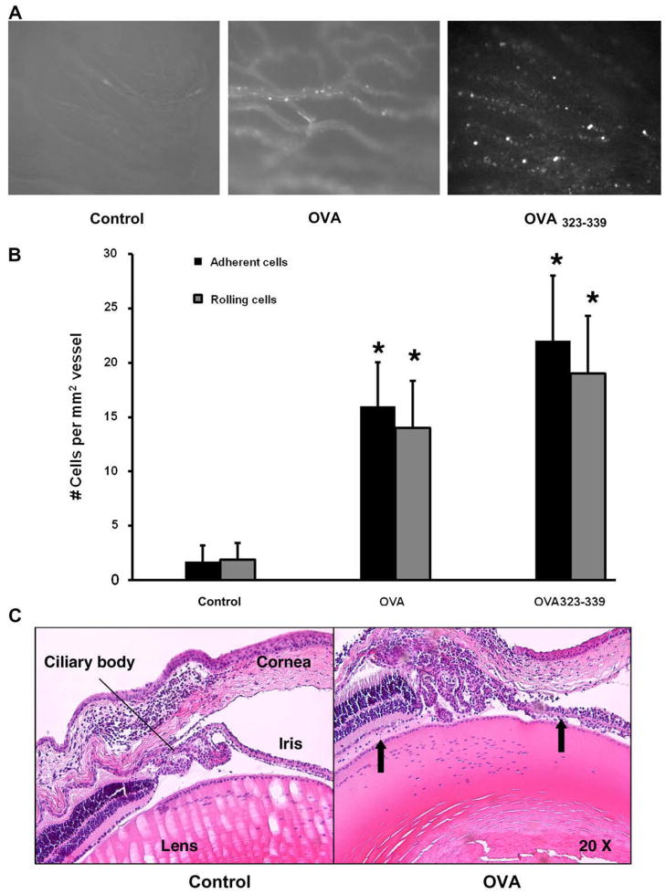
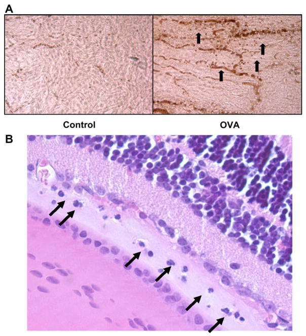
T cell-mediated uveitis is strongly associated with many systemic inflammatory disorders. Th17 cells are a novel T cell subset characterized by production of interleukin (IL)-17. In this study, we used DO11.10 mice to investigate the role of IL-17 in the pathogenesis of uveitis. CD4(+) T cells in DO11.10 mice are genetically engineered to react with ovalbumin (OVA). IL-17 expression was determined by real-time PCR and ELISPOT. Uveitis was induced by intravitreal injection of OVA, and ocular inflammation was evaluated by intravital microscopy. OVA challenge significantly induced IL-17 production by DO11.10 splenocytes in vitro. Next, we examined whether OVA challenge could elicit local inflammation and induce IL-17 in vivo. OVA elicited marked neutrophil-predominant inflammatory cell infiltration in the eyes. This leukocyte influx was mediated by CD4(+) lymphocytes as evidenced by significant inhibition of the ocular inflammation by CD4+ depleting antibody. Compared to control mice, OVA treatment induced IL-17 expression. Moreover, anti-IL-17 antibody markedly reduced OVA-mediated ocular inflammation. Finally, the neutralization of IL-17 attenuated ocular expression of CXCL2 and CXCL5, two cytokines which are chemotactic for neutrophils. Our study suggests that IL-17 is implicated in the pathogenesis of this T cell-mediated model of uveitis in part through neutrophil chemotaxis as a downstream effect of IL-17.


Figures

Fig. 1
Fig. 1
OVA specifically induces IL-17 production in the splenocytes from DO11.10 mice. Splenocytes were isolated from DO11.10 and BALB/c mice. They were divided into 3 groups (n = 5): Control, OVA, and anti-CD3 and anti-CD28 antibodies. After 24-h stimulation, IL-17 production was measured by ELISPOT. IL-17 activity is defined as number of spots per 400,000 input cells (*p < 0.01).
Fig. 2
Fig. 2
OVA induces IL-17 production in a time-dependent manner. Splenocytes were isolated from DO11.10 mice. These cells were further stimulated with OVA for 3, 9, 21, 27, 33, 45 and 51 h, respectively (n = 3). IL-17 production was determined by ELISPOT.
Fig. 3
Fig. 3
Intravitreal injection of OVA elicits uveitis in DO11.10 mice. OVA or PBS as a control vehicle was administered intravitreally (n = 12). In addition, a group of mice received intravitreal injection of OVA323–339 peptide (n = 4). Twenty-four hours later, circulating leukocytes were labeled with rhodamine by intraperitoneal injection, and ocular inflammation was assessed by intravital microscopy. (A) Representative image of a single frame from intravital microscopy videos taken of the iris at 24 h during the course of inflammation induced by OVA. Note: Influx and adhesion of rhodamine-labeled leukocytes in OVA-challenged eye. (B) Quantization of rolling and adherent cells in the vascular bed of the iris (*p < 0.05). (C) Representative histology of mouse eyes challenged with and without OVA. The ocular tissue was harvested for H&E staining. The inflammatory change was evaluated by light microscopy. Anterior chamber inflammation and a leukocytic infiltrate posterior to the ciliary body and exudates (arrows) were observed in the eye treated with OVA compared to control group.
Fig. 4
Fig. 4
Predominant neutrophil infiltration occurs in the eye of DO11.10 mice at 24 h after OVA challenge. OVA or PBS as a control vehicle was administered intravitreally (n = 4). (A) Twenty-four hours later, the eyes were enucleated, dissected, fixed in paraformaldehyde and the anterior segment was incubated with Ly6G antibody. Note: Majority of infiltrating cells were Ly6G positive (arrows). (B) Further histology showed that infiltrating leukocytes were mainly polymorphonuclear neutrophils (arrows).
Fig. 5
Fig. 5
Depletion of CD4+ cells inhibits OVA-induced uveitis in DO11.10 mice. DO11.10 mice received 100 μg of GK1.5 antibody intraperitoneally for consecutive 2 days to deplete peripheral CD4+ cells (n = 3). Then, OVA was delivered intravitreally to induce uveitis. Twenty-four hours later, ocular inflammatory cells were assessed by intravital microscopy. (A) Representative image of a single frame from intravital microscopy videos taken of the iris at 24 h during the course of inflammation induced by OVA. Note: GK1.5 antibody prevented OVA-induced inflammatory cell infiltration in the eye. (B) Quantization of rolling and adherent cells in the vasculature of the iris treated with and without GK1.5 antibody (*p < 0.05).
Fig. 6
Fig. 6
Interferon-γ neutralizing antibody does not inhibit OVA-induced ocular inflammation in DO11.10 mice. Anti-interferon-γ antibody (15 μg per eye) was injected intravitreally along with OVA into the eye of DO11.10 mice (n = 4). Twenty-four hours later, ocular inflammatory cells were assessed by intravital microscopy. Then, total RNA was harvested for RT-PCR. (A) Representative image of a single frame from intravital microscopy videos taken of the iris at 24 h during the course of inflammation induced by OVA. Note: Anti-interferon-γ antibody did not suppress OVA-induced inflammatory cell infiltration in the eye. (B) Quantization of rolling and adherent cells in the vasculature of the iris treated with and without anti-interferon-γ antibody.
Fig. 7
Fig. 7
OVA induces IL-17 gene expression in the eye of DO11.10 mice. OVA or PBS as a control vehicle was administered intravitreally. Twenty-four hours later, the eyes were harvested and total RNA was collected. RT-PCR analysis revealed upregulation of IL-17 in OVA-treated eyes (n = 6).
Fig. 8
Fig. 8
Infiltration of IL-17+ cells occurs in the eye of DO11.10 mice at 24 h after OVA challenge. OVA or PBS as a control vehicle was administered intravitreally (n = 3). Twenty-four hours later, the iris was dissected and fixed in paraformaldehyde. Immunohistochemical analysis was performed to detect IL-17 protein expression in the eye. Note: IL-17+ cells (arrows) were only detected in OVA-treated eye but not in the control group.
Fig. 9
Fig. 9
Infiltrating CD4+ T cells express IL-17 in the eye of DO11.10 mice at 24 h after OVA challenge. OVA or PBS as a control vehicle was administered intravitreally (n = 4). Twenty-four hours after OVA stimulation, the iris was dissected, and fixed in paraformaldehyde. Two-color immunofluorescent staining was used to determine CD4 (FITC), green color: A) and IL-17 (APC, red color: B). The expression of CD4 and IL-17 was further analyzed by co-localization of both fluorescence colors (C). Note: Most IL-17-expressed cells were CD4+ lymphocytes.
Fig. 10
Fig. 10
Depletion of NKT cells does not inhibit OVA-induced uveitis in DO11.10 mice. DO11.10 mice received 200 μg of anti-CD161 antibody intraperitoneally twice 3 days apart before uveitis induction (n = 3). Then, OVA was delivered intravitreally to induce uveitis. Twenty-four hours later, ocular inflammatory cells were assessed by intravital microscopy. (A) Representative image of a single frame from intravital microscopy videos taken of the iris at 24 h during the course of inflammation induced by OVA. Note: anti-CD161 antibody did not reduce OVA-induced inflammatory cell infiltration in the eye. (B) Quantization of rolling and adherent cells in the vasculature of the iris treated with and without anti-CD161 antibody.
Fig. 11
Fig. 11
IL-23p19 neutralizing antibody did not inhibit OVA-induced ocular inflammation in DO11.10 mice. Anti-IL-23p19 antibody (100 μg per mouse) was injected intraperitoneally into DO11.10 mice when they received intravitreal OVA challenge (n = 3). Twenty-four hours later, ocular inflammatory cells were assessed by intravital microscopy. (A) Representative image of a single frame from intravital microscopy videos taken of the iris at 24 h during the course of inflammation induced by OVA. Note: Anti-IL-23p19 antibody did not suppress OVA-induced inflammatory cell infiltration in the eye. (B) Quantization of rolling and adherent cells in the vasculature of the iris treated with and without anti-IL-23p19 antibody.
Fig. 12
Fig. 12
Anti-IL-17 antibody inhibits OVA-induced ocular inflammation in DO11.10 mice. Anti-IL-17 antibody (20 μg per eye) was injected intravitreally along with OVA into the eye of DO11.10 mice (n = 7). Twenty four hours later, ocular inflammatory cells were assessed by intravital microscopy. Then, total RNA was harvested for RT-PCR. (A) Representative image of a single frame from intravital microscopy videos taken of the iris at 24 h during the course of inflammation induced by OVA. Note: Anti-IL-17 antibody inhibited OVA-induced inflammatory cell infiltration in the eye. (B) Quantitation of rolling and adherent cells in the vasculature of the iris treated with and without anti-IL-17 antibody (*p < 0.05). (C) RT-PCR analysis of CXCL2 and CXCL5 revealed up-regulation of these 2 chemokines in the eyes treated with OVA. This up-regulation was reduced by anti-IL-17 antibody.

Similar articles

Cited by

References

    1. Gritz DC, Wong IG. Incidence and prevalence of uveitis in Northern California; the Northern California Epidemiology of Uveitis Study. Ophthalmology. 2004;111(3):491–500. discussion 500. - PubMed
    1. Nussenblatt RB. The natural history of uveitis. International Ophthalmology. 1990;14(5–6):303–8. - PubMed
    1. Becker MD, Adamus G, Davey MP, Rosenbaum JT. The role of T cells in autoimmune uveitis. Ocular Immunology & Inflammation. 2000;8(2):93–100. - PubMed
    1. Martin TM, Smith JR, Rosenbaum JT. Anterior uveitis: current concepts of pathogenesis and interactions with the spondyloarthropathies. Current Opinion in Rheumatology. 2002;14(4):337–41. - PubMed
    1. Atalla L, Linker-Israeli M, Steinman L, Rao NA. Inhibition of autoimmune uveitis by anti-CD4 antibody. Investigative Ophthalmology & Visual Science. 1990;31(7):1264–70. - PubMed