. 2009 May;198(1):1-7.
doi: 10.1016/j.jmr.2008.12.007. Epub 2008 Dec 14.

Determining the helical tilt of membrane peptides using electron paramagnetic resonance spectroscopy

Affiliations

Determining the helical tilt of membrane peptides using electron paramagnetic resonance spectroscopy

Justin P Newstadt et al. J Magn Reson. 2009 May.

Abstract

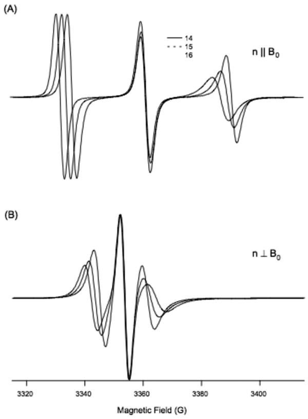
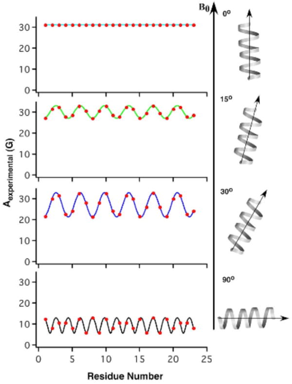
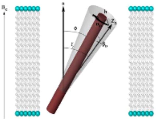
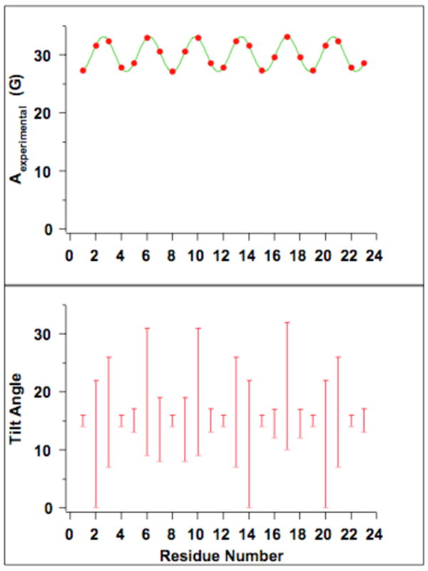
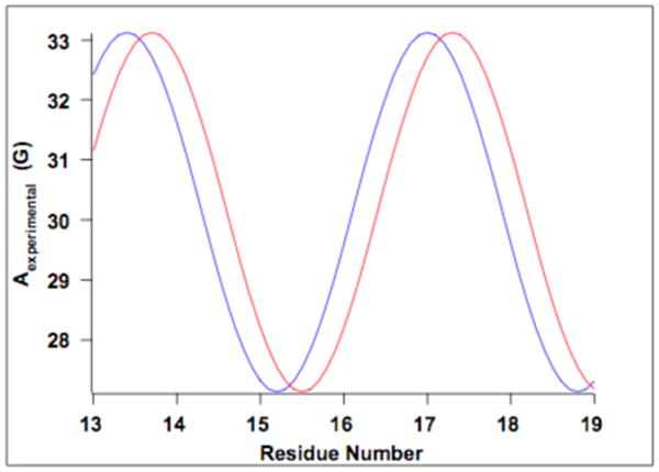
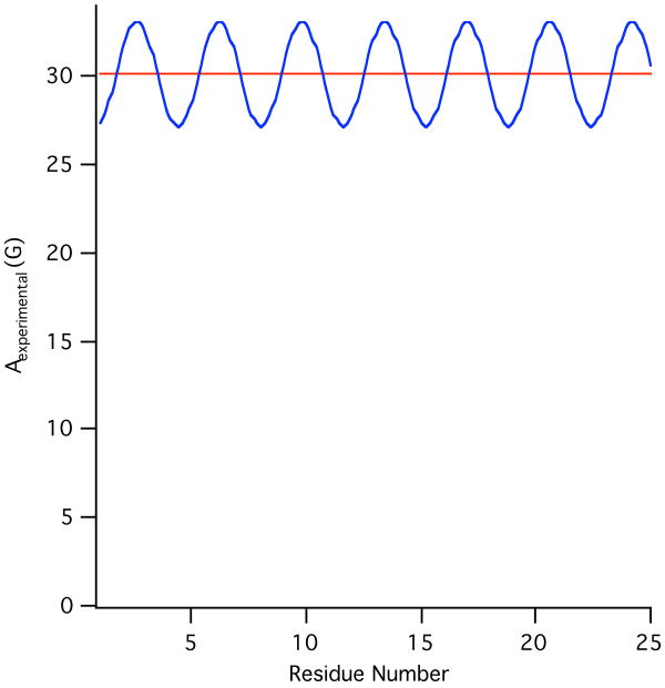
Theoretical calculations of hyperfine splitting values derived from the EPR spectra of TOAC spin-labeled rigid aligned alpha-helical membrane peptides reveal a unique periodic variation. In the absence of helical motion, a plot of the corresponding hyperfine splitting values as a function of residue number results in a sinusoidal curve that depends on the helical tilt angle that the peptide makes with respect to the magnetic field. Motion about the long helical axis reduces the amplitude of the curve and averages out the corresponding hyperfine splitting values. The corresponding spectra can be used to determine the director axis tilt angle from the TOAC spin label, which can be used to calculate the helical tilt angle due to the rigidity of the TOAC spin label. Additionally, this paper describes a method to experimentally determine this helical tilt angle from the hyperfine splitting values of three consecutive residues.


Figures

Figure 1
Figure 1
Simulation of EPR spectra for residues 14-16 demonstrating how the Aexp values change between these residues. (A) Parallel aligned (B) perpendicularly aligned with respect to the direction of magnetic field (B0).
Figure 2
Figure 2
Hyperfine waves as a function of helical tilt angle, using predefined values for A (33.3 G), A (5.6 G), βD (21°), and αD at residue 18 (-10°).
Figure 3
Figure 3
Illustration of an α-helix with attached TOAC spin label and applied magnetic field. φ represents the helical tilt angle, ζ, (equal to ψ in the parallel orientation) is the director tilt angle, which is directly measured in the EPR spectrum. ZD is the director axis of the spin label and to the axis directed along the π-orbital, perpendicular to the N-O bond of the TOAC spin label (not shown). αD is the rotation of the spin label around the helix and depends on the residue number, while βD is the angle between the director axis and the helical axis.
Figure 4
Figure 4
Helical tilt ranges as a function of residue number. Using simulated Aexp values corresponding to a tilt angle of 15°, helical tilt ranges were found. As can be seen in the figure, some residues result in helical tilt ranges that are much smaller than others, which is due to changes in the residues' orientation with respect to the magnetic field (changing values of αD). Furthermore, a relatively narrow helical tilt range is found within three consecutive residues.
Figure 5
Figure 5
Hyperfine wave as a function of reside number and showing a shift in the hyperfine waves due to changing the αD frame. The blue wave represents the correct αD frame, while the red wave is out of phase by 30°. When the wave is out of phase, it shifts either to the right or to the left and causes the corresponding helical tilt angle ranges to become incompatible with each other as a result, which can be seen in Table 3.
Figure 6
Figure 6
Illustration of the effects of static (blue) and fast rotational dynamics (red) about the long helical axis of the peptide aligned in a membrane. Using simulated Aexp values corresponding to a tilt angle of 15°, βD (21°), A (33.3 G), A (5.6 G), αD (-10). Rapid increase of axial rotation about the long helical axis of the peptide causes an averaging of the amplitude of the hyperfine waves as dictated by equation 4.

Similar articles

Cited by

References

    1. Mesleh MF, Lee S, Veglia G, Thiriot DS, Marassi FM, Opella SJ. Dipolar waves map the structure and topology of helices in membrane proteins. J Am Chem Soc. 2003;125:8928–8935. - PMC - PubMed
    1. Arkin IT, MacKenzie KR, Fisher L, Aimoto S, Engelman DM, Smith SO. Mapping the lipid-exposed surfaces of membrane proteins. Nat Struct Biol. 1996;3:240–243. - PubMed
    1. Nagy JK, Lau FW, Bowie JU, Sanders CR. Mapping the oligomeric interface of diacylglycerol kinase by engineered thiol cross-linking: Homologous sites in the transmembrane domain. Biochemistry. 2000;39:4154–4164. - PubMed
    1. Doyle DA, Cabral JM, Pfuetzner RA, Kuo AL, Gulbis JM, Cohen SL, Chait BT, MacKinnon R. The structure of the potassium channel: Molecular basis of K+ conduction and selectivity. Science. 1998;280:69–77. - PubMed
    1. Fu DX, Libson A, Miercke LJW, Weitzman C, Nollert P, Krucinski J, Stroud RM. Structure of a glycerol-conducting channel and the basis for its selectivity. Science. 2000;290:481–486. - PubMed

Publication types

LinkOut - more resources