

Review
doi: 10.1016/j.jfms.2008.09.008.
Epub 2009 Feb 28.
A review of feline infectious peritonitis virus infection: 1963-2008
Affiliations
- PMID: 19254859
- PMCID: PMC7129802
- DOI: 10.1016/j.jfms.2008.09.008
Item in Clipboard
Review
A review of feline infectious peritonitis virus infection: 1963-2008
J Feline Med Surg.
2009 Apr.
No abstract available
Figures
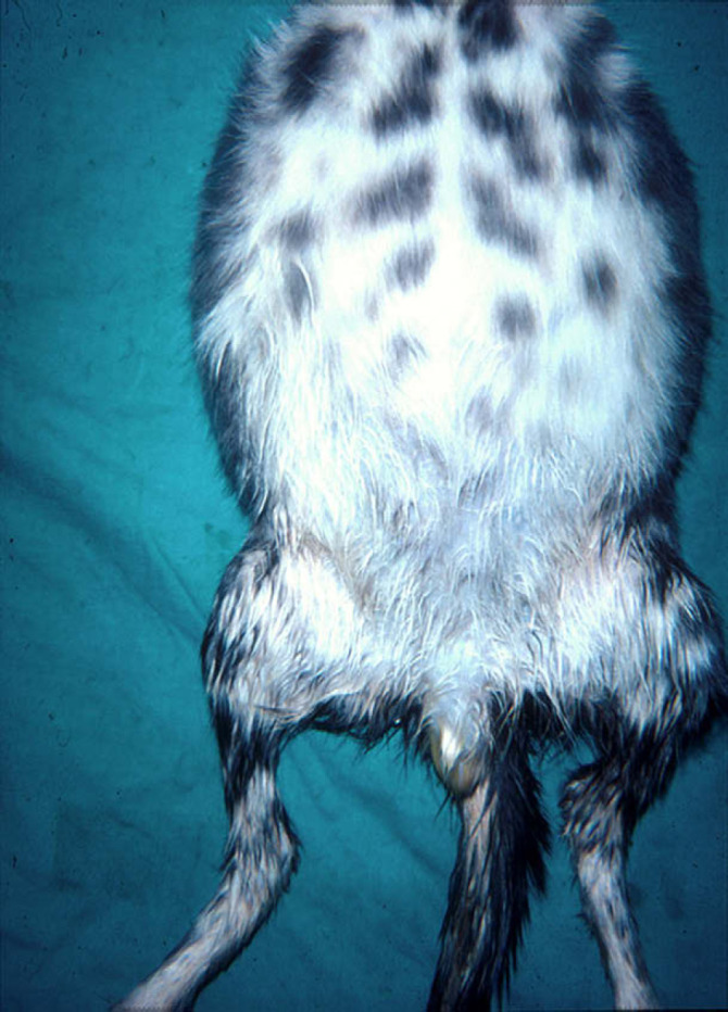
Grossly distended abdomen of a kitten with effusive FIP. Note the scrotal enlargement due to inflammation of the tunics.
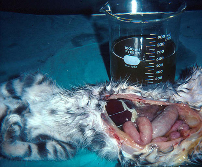
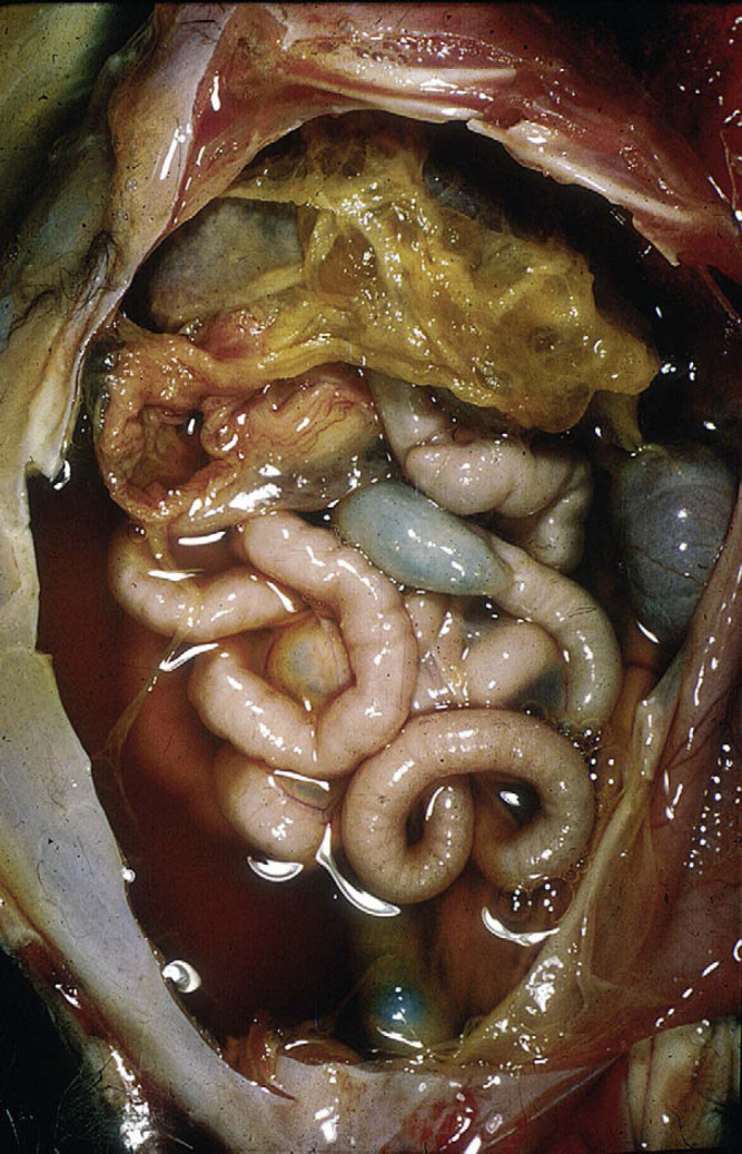
Over 600 ml of a yellow, mucinous effusion was removed from the abdomen at necropsy. Note fibrin tags on liver and spleen and ground glass appearance of the serosa.
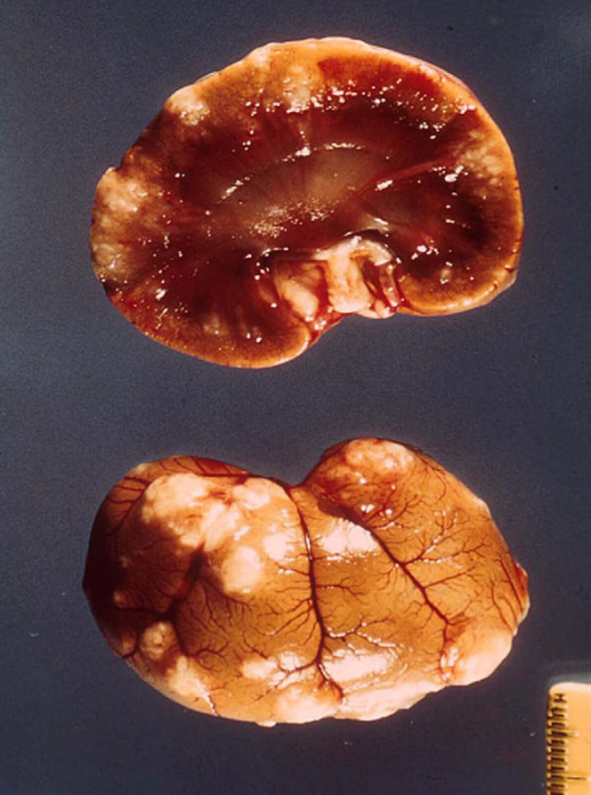
Cross section of a kidney from a cat with dry FIP. Numerous granulomatous lesions are seen on the capuslue of the kidney and extending downward into the parenchyma.
Enlarged mesenteric lymph node in a cat with the dry form of FIP. Note the residual fibrinous plaque on the spleen. Such residual lesions support the concept that many cases of dry FIP began as a brief bout of wet FIP.
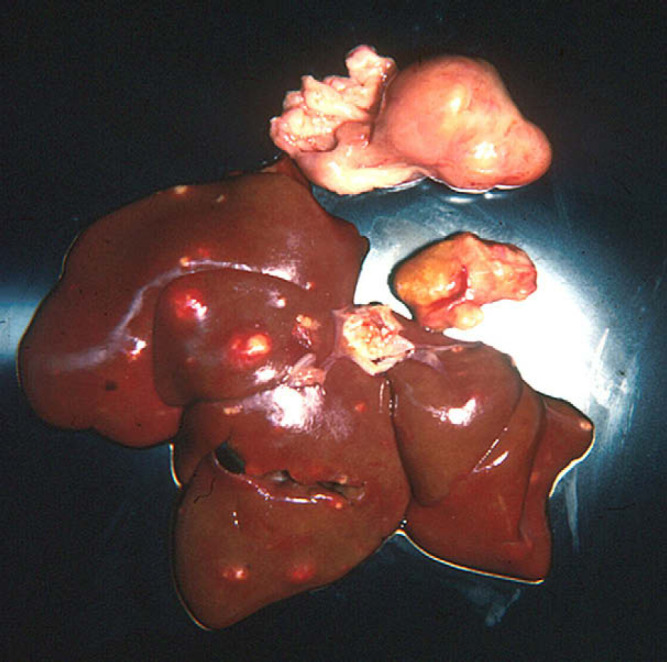
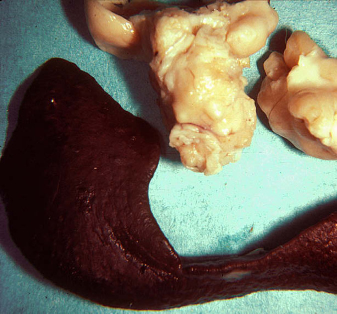
Mesenteric and hepatic lymph nodes and liver from a cat with non-effusive FIP. The lymph nodes are enlarged and involved with granulomatous adenitis. The liver capsule contains raised, whitish foci 0.5–1 cm in diameter, extending into the underlying parenchyma.
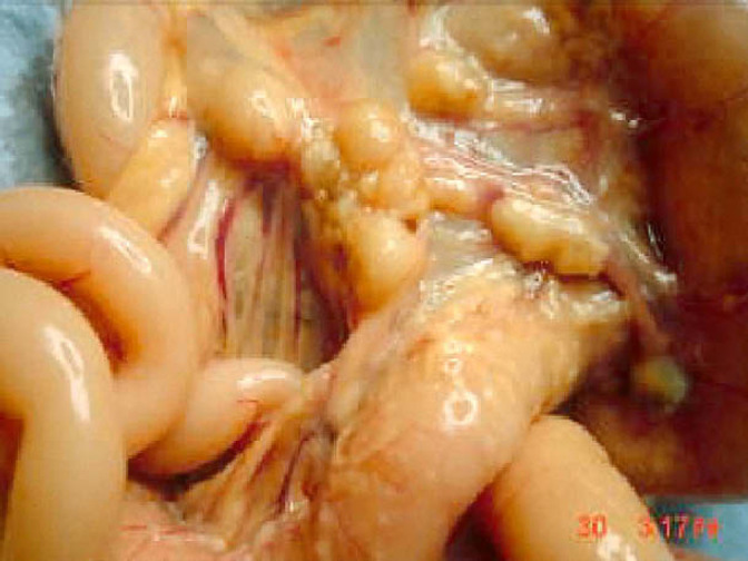
Gross appearance of the caecum, colon and ceco-colic lymph nodes of a cat with the intestinal form of dry FIP.
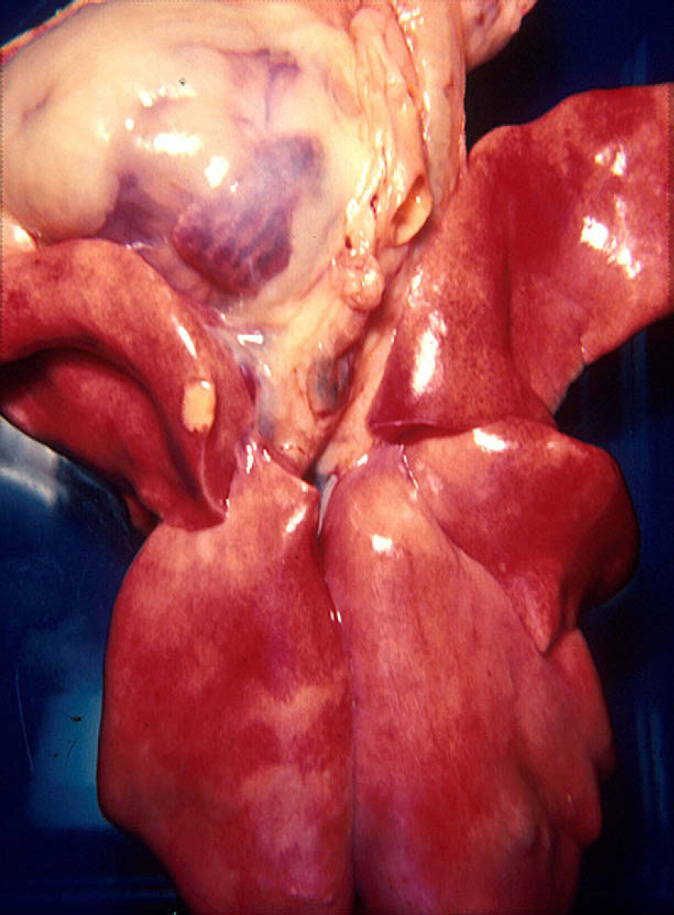
Lungs and heart of cat shown in Fig 5. A solitary pleural granuloma is noted along the edge of a cranial lung lobe.

Uveitis of the right eye in a cat with the dry form of FIP. The color of the iris has changed, the anterior chamber is somewhat hazy, and there is a pigmented lesion in the center of the cornea (a keratic precipitate). Note the irregularity in the shape of the right pupil compared to the normal left pupil.
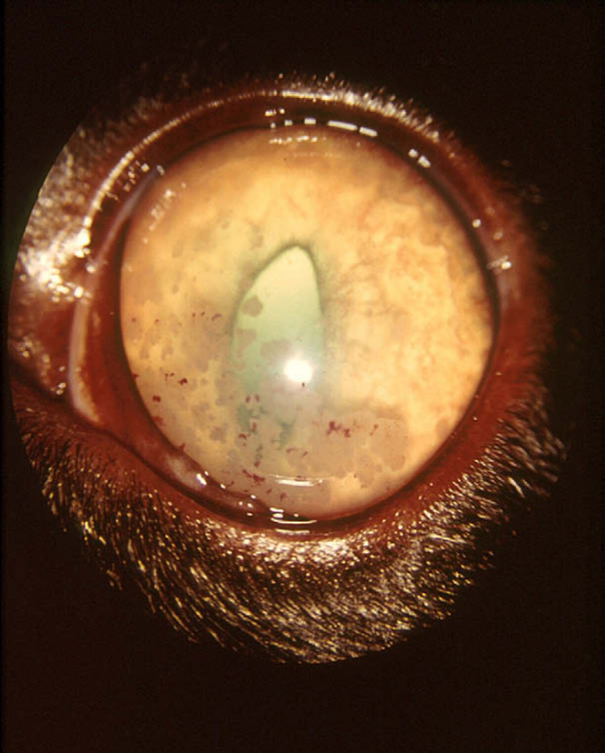
Keratic precipitates on the inner cornea of a cat with dry FIP. Note the reversed D-shape of pupil due to infiltration of the iris.
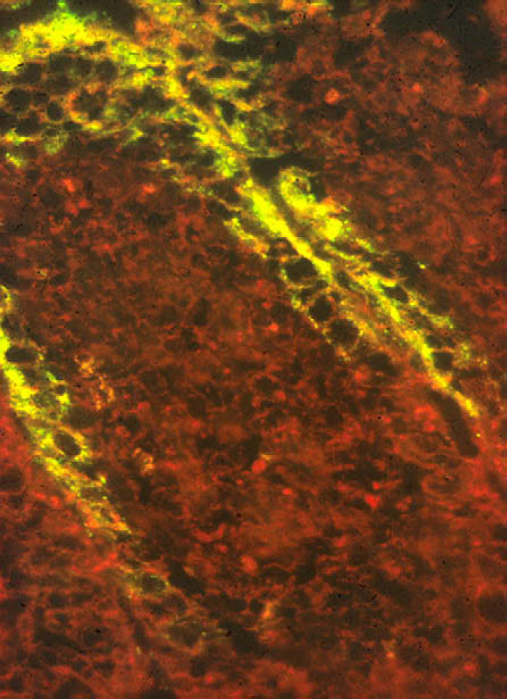
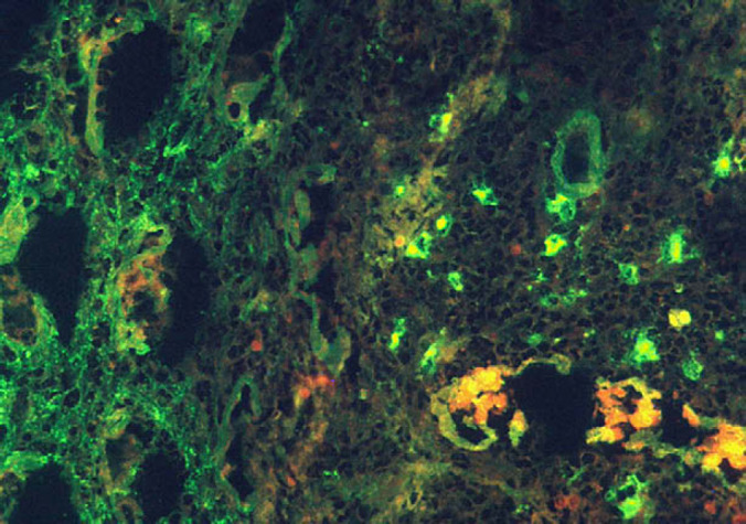
Immunofluorescent antibody staining for feline coronavirus antigens on a section of small intestine from a cat experimentally infected with FIPV. Virus is concentrated in the mature apical epithelium at the tips of the intestinal villi.
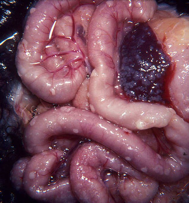
Abdominal viscera of a cat with effusive FIP. The serosal surface of the intestines and spleen are covered with punctate, coalescing fibrinous plaques, the classic pyogranulomas of effusive FIP. Some peritoneal effusion is evident, although most has been removed.
Abdominal viscera of a cat with effusive FIP. The serosal surface of the intestines and spleen is covered with punctate, coalescing fibrinous plaques, the classic pyogranulomas of effusive FIP. Some peritoneal effusion remains, though most has been removed.
Abdominal viscera of a cat with effusive FIP. The serosal surface of the intestines and spleen is covered with punctate, coalescing fibrinous plaques, the classic pyogranulomas of effusive FIP. Some peritoneal effusion remains, though most has been removed.
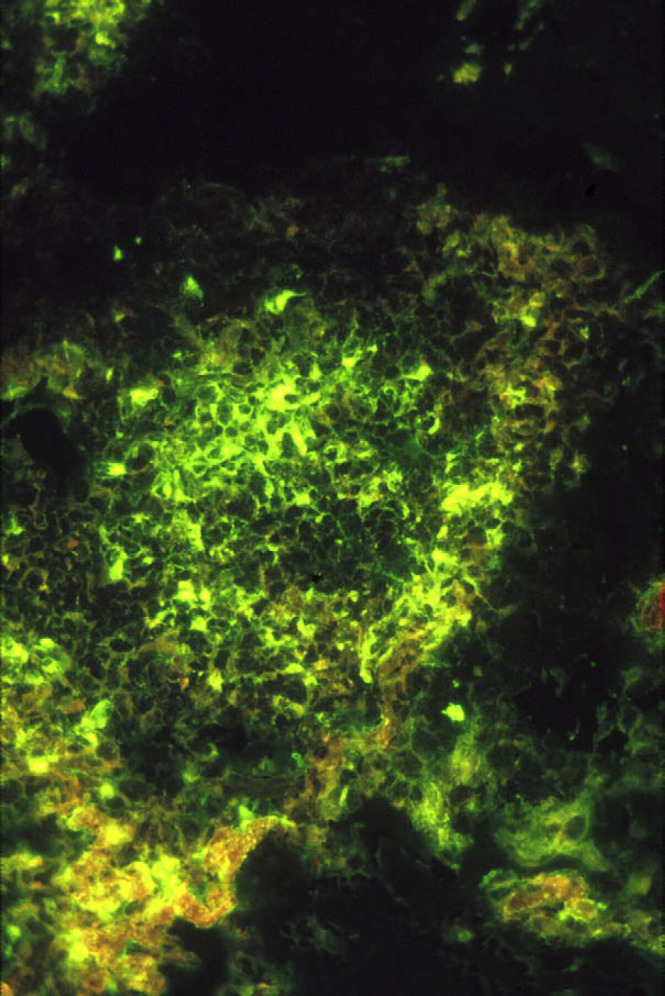
Immunofluorescent antibody staining for feline coronavirus antigen of a pyogranuloma in the serosa of the bladder. Antigen is concentrated in a high proportion of the macrophages within focal pyogranulomas.
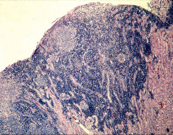
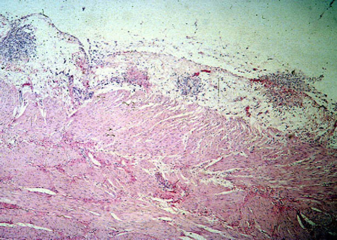
Microscopic view of a granulomatous lesion from a cat with non-effusive FIP. Distinct focal accumulations of macrophages are seen just under the capsule of the kidney. These foci are surrounded by dense accumulations of lymphocytes and plasma cells extending downward into the parenchyma.
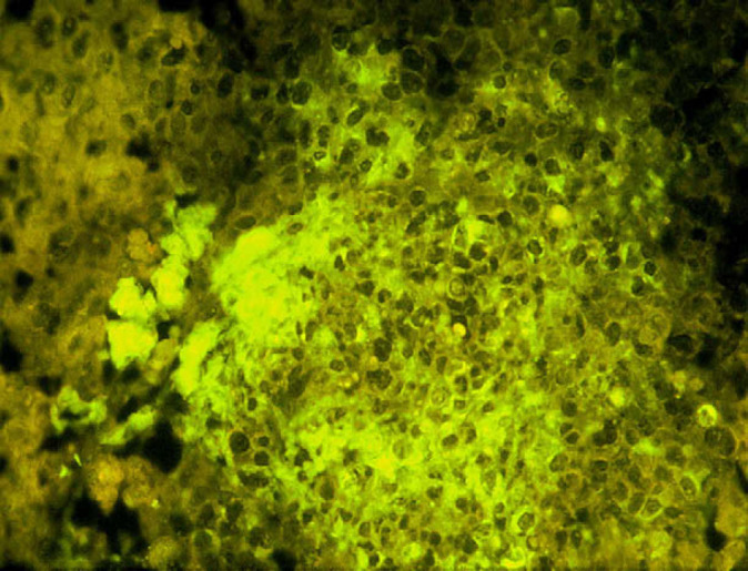
Immunofluorescent staining of a foci from the kidney section shown in Fig 15. A much smaller porportion of the macrophages stain for feline coronavirus antigen than in the pyrogranulomas of effusive FIP (Fig 14).
Immunofluorescent antibody staining for feline IgG. Accumulations of IgG are seen both within and without macrophages, mainly within and adjacent to pyrogranulomas. Some plasma cells also contain IgG.
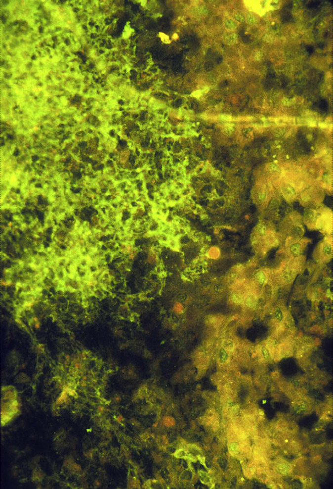
Immunofluorescent antibody staining for feline C3. Complement staining of macrophages has a web-like appearance.
References
-
- Holzworth J.E. Some important disorders of cats, Cornell Vet 53, 1963, 157–160. - PubMed
-
- Feldman B.F., Jortner B.S. Clinico-pathology conference, J Am Vet Med Assoc 144, 1965, 1409–1411. - PubMed
-
- Wolfe L.G., Griesemer R.A. Feline infectious peritonitis, Pathol Vet 3, 1966, 255–270. - PubMed
-
- Zook B.C., King N.W., Robinson R.L., McCombs H.L. Ultrastructural evidence for the viral etiology of feline infectious peritonitis, Pathol Vet 5, 1968, 91–95.
Publication types
MeSH terms
Substances
LinkOut - more resources
Full Text Sources
Other Literature Sources
