. 2009 May;37(8):2449-60.
doi: 10.1093/nar/gkp101. Epub 2009 Mar 2.

The chromatin remodeling factor CHD8 interacts with elongating RNA polymerase II and controls expression of the cyclin E2 gene

Affiliations

The chromatin remodeling factor CHD8 interacts with elongating RNA polymerase II and controls expression of the cyclin E2 gene

M Rodríguez-Paredes et al. Nucleic Acids Res. 2009 May.

Abstract

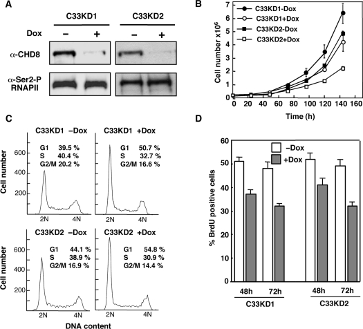
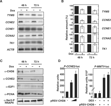
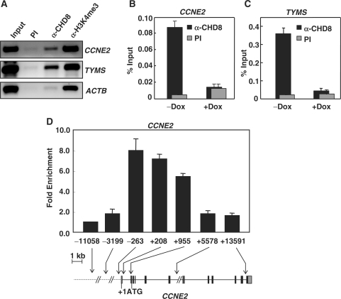
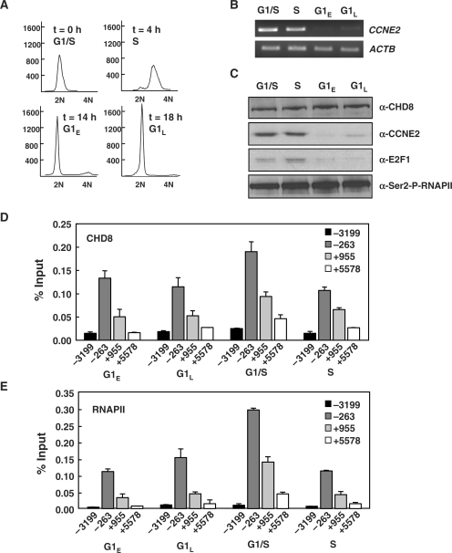
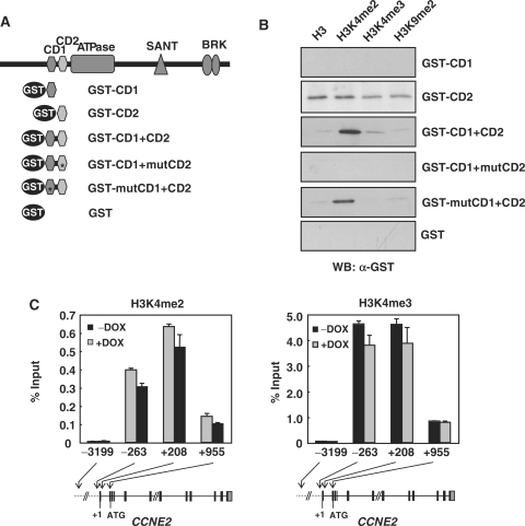
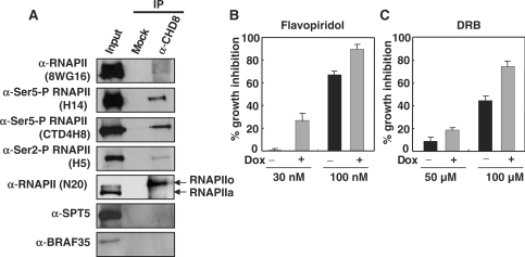
CHD8 is a chromatin remodeling ATPase of the SNF2 family. We found that depletion of CHD8 impairs cell proliferation. In order to identify CHD8 target genes, we performed a transcriptomic analysis of CHD8-depleted cells, finding out that CHD8 controls the expression of cyclin E2 (CCNE2) and thymidylate synthetase (TYMS), two genes expressed in the G1/S transition of the cell cycle. CHD8 was also able to co-activate the CCNE2 promoter in transient transfection experiments. Chromatin immunoprecipitation experiments demonstrated that CHD8 binds directly to the 5' region of both CCNE2 and TYMS genes. Interestingly, both RNA polymerase II (RNAPII) and CHD8 bind constitutively to the 5' promoter-proximal region of CCNE2, regardless of the cell-cycle phase and, therefore, of the expression of CCNE2. The tandem chromodomains of CHD8 bind in vitro specifically to histone H3 di-methylated at lysine 4. However, CHD8 depletion does not affect the methylation levels of this residue. We also show that CHD8 associates with the elongating form of RNAPII, which is phosphorylated in its carboxy-terminal domain (CTD). Furthermore, CHD8-depleted cells are hypersensitive to drugs that inhibit RNAPII phosphorylation at serine 2, suggesting that CHD8 is required for an early step of the RNAPII transcription cycle.


Figures

Figure 1.
Figure 1.
Depletion of CHD8 impairs cell proliferation. CHD8 shRNA knockdown cell lines (C33KD1 and C33KD2) were generated by stable transfection of C33A cells. (A) Immunoblot analysis of Dox-treated (+) and non-treated (−) cells after 72 h, using α-CHD8 antibody. α-Ser2-P RNAPII antibody (H5) was used as a loading control. (B) Growth curve of CHD8 knockdown cell lines in the presence or absence of Dox. Data are the average of three independent experiments. Error bars represent standard deviation. (C) Flow cytometry analysis of asynchronous C33KD1 and C33KD2 cells treated with or without Dox for 72 h. Representative experiments are shown. Numbers are the average of three independent experiments. (D) BrdU incorporation analysis of cycling cells. C33KD1 and C33KD2 cells treated or non-treated with Dox for 48 and 72 h were labeled with BrdU as described in Methods. Percentage of cells that had incorporated BrdU was determined by immunofluorescence using α-BrdU antibodies. Numbers are the average of two independent experiments.
Figure 2.
Figure 2.
Depletion of CHD8 leads to down-regulation of TYMS and CCNE2 genes. (A) Transcript levels of genes induced at the G1/S transition (TYMS, CCNE2, CCNE1, CCNA2, TK1) were analyzed by RT–PCR in Dox-treated (+) or non-treated (−) C33KD2 cells, at the indicated times. β-Actin (ACTB) transcript levels were also determined as a control for the amount of input cDNA. (B) Quantification of signals shown in (A). Values were normalized to the ACTB gene transcript level. The level of cDNA amplified in Dox non-treated cells (−) was considered 100%. Values are the average of three independent experiments. Bars indicate standard error of the mean. (C) Levels of CHD8, CCNE2, E2F1 and PCNA proteins were analyzed by immunoblot in C33KD2 cells treated (+) or non-treated (−) with Dox during the indicated time. α-Ser2-P-RNAPII antibody (H5) was used as a loading control. (D) The CCNE2 promoter is activated by CHD8. COS-7 cells were transfected with 0.2 µg of pCycE2-LUC plasmid along with 0.3 or 0.7 µg of pRSV-CHD8 plasmid. 0.2 µg of the pMMTV-LUC plasmid were co-transfected with a glucocorticoid receptor expression vector (0.2 µg) and with or without 0.7 µg of pRSV-CHD8. When indicated, the glucocorticoid receptor ligand dexamethasone (DEX) was added at 1 µg/ml for 12 h. Values are the average of three independent experiments ± SD.
Figure 3.
Figure 3.
Analysis of the presence of CHD8 in the 5′ region of the CCNE2 and TYMS genes by ChIP. (A) Crosslinked DNA–protein complexes from C33A cells were immunoprecipitated with α-CHD8 and α-H3K4me3 (as a positive control). PCR amplification was performed with specific primers of the CCNE2, TYMS and ACTB genes. The center of PCR products relative to the transcription start sites is as follows: +836 for the CCNE2, +91 for the TYMS and +1294 for the ACTB genes. (B and C) Binding of CHD8 to the CCNE2 (B) and TYMS (C) genes in C33KD2 cells treated or not with Dox for 72 h. (D) Distribution of CHD8 over the CCNE2 gene in C33A cells. Numbers correspond to the center nucleotide of the real time PCR product relative to the transcription start site. A diagram of the CCNE2 gene is also shown. Black boxes correspond to coding exons while grey boxes correspond to the 5′UTR and 3′UTR. Transcription start point (+1) and ATG are also indicated. In (B), (C) and (D), immunoprecipitated DNA from three independent experiments was quantified using qPCR and expressed as percentage of the input (B and C) or as fold enrichment respect to the −11 058 amplicon (D). PI, pre-immune serum.
Figure 4.
Figure 4.
CHD8 and RNAPII bind to CCNE2 promoter-proximal region regardless of the cell-cycle phase. (A) HeLa cells were synchronized by double thymidine blocking as described in ‘Materials and Methods’ section. After the release of the blockade (t = 0), samples were taken at the indicated times for flow cytometry analysis. Synchronized cells were taken at the indicated times and levels of CCNE2 and ACTB transcripts (B) and CHD8, Cyclin E2 and E2F1 proteins (C) were determined by RT–PCR and immunoblot analysis, respectively. α-E2F1 was used as a control of a typical G1/S- and S-expressed gene. α-Ser2-P-RNAPII antibody (H5) was used as a loading control for immunoblotting experiments. (D and E) ChIP analysis of the binding of CHD8 (D) and total RNAPII (N20 antibody) (E) to the CCNE2 gene in different cell-cycle phases. Numbers correspond to the center nucleotide of the real-time PCR product relative to the transcription start site. Data are the average of 12 qPCR determinations from two independent experiments.
Figure 5.
Figure 5.
The tandem chromodomains of CHD8 bind to H3K4me2, in vitro. (A) Schematic of the CHD8 fragments used for binding studies. (B) Purified GST, GST-CD1, GST-CD2, GST-CD1+CD2, GST-CD1+mutCD2 and GST-mutCD1+CD2 proteins were incubated with different biotinylated histone H3 N-terminal peptides, such as unmodified (H3), Lys 4-di-methylated (H3K4me2), Lys 4-tri-methylated (H3K4me3) or Lys 9-di-methylated (H3K9me2). Bound fractions from the binding reactions were analyzed by western blotting using α-GST antibody. A representative experiment is shown. (C) Analysis of the levels of H3K4me2 and H3K4me3 in the 5′ region of the CCNE2 gene by ChIP in C33KD2 cells treated or not with Dox during 72 h. Immunoprecipitated DNA from three independent experiments was quantified using qPCR. Numbers correspond to the center nucleotide of the real time PCR product relative to the transcription start site. Data are expressed as percentage of the input.
Figure 6.
Figure 6.
CHD8 interacts with elongating RNAPII. (A) CHD8 associates with RNAPIIo in vivo. Immunoblot analysis of C33A total extract (input) and fractions immunoprecipitated with α-CHD8 or nonspecific antiserum (mock). Antibodies used for immunoblot are indicated on the left in each panel. (B and C) CHD8-depleted cells are hypersensitive to transcription elongation inhibitors. C33KD2 cells were grown with or without Dox and with or without flavopiridol (B) or DRB (C) at the indicated concentrations. Total cell numbers were determined after 5 days under the indicated conditions. Percentage of growth inhibition in the presence of either of flavopiridol (B) or DRB (C) is represented.

Similar articles

Cited by

References

    1. Saunders A, Core LJ, Lis JT. Breaking barriers to transcription elongation. Nat. Rev. Mol. Cell Biol. 2006;7:557–567. - PubMed
    1. Li B, Carey M, Workman JL. The role of chromatin during transcription. Cell. 2007;128:707–719. - PubMed
    1. Smith CL, Peterson CL. ATP-dependent chromatin remodeling. Curr. Top. Dev. Biol. 2005;65:115–148. - PubMed
    1. Flaus A, Owen-Hughes T. Mechanisms for ATP-dependent chromatin remodelling: farewell to the tuna-can octamer? Curr. Opin. Genet. Dev. 2004;14:165–173. - PubMed
    1. Flaus A, Martin DM, Barton GJ, Owen-Hughes T. Identification of multiple distinct Snf2 subfamilies with conserved structural motifs. Nucleic Acids Res. 2006;34:2887–2905. - PMC - PubMed

Publication types

MeSH terms