. 2009 May;29(9):2372-89.
doi: 10.1128/MCB.01505-08. Epub 2009 Mar 2.

Similar and distinct properties of MUPP1 and Patj, two homologous PDZ domain-containing tight-junction proteins

Affiliations

Similar and distinct properties of MUPP1 and Patj, two homologous PDZ domain-containing tight-junction proteins

Makoto Adachi et al. Mol Cell Biol. 2009 May.

Abstract

MUPP1 and Patj are both composed of an L27 domain and multiple PDZ domains (13 and 10 domains, respectively) and are localized to tight junctions (TJs) in epithelial cells. Although Patj is known to be responsible for the organization of TJs and epithelial polarity, characterization of MUPP1 is lacking. In this study, we found that MUPP1 and Patj share several binding partners, including JAM1, ZO-3, Pals1, Par6, and nectins (cell-cell adhesion molecules at adherens junctions). MUPP1 and Patj exhibited similar subcellular distributions, and the mechanisms with which they localize to TJs also appear to overlap. Despite these similarities, functional studies have revealed that Patj is indispensable for the establishment of TJs and epithelial polarization, whereas MUPP1 is not. Thus, although MUPP1 and Patj share several molecular properties, their functions are entirely different. We present evidence that the signaling mediated by Pals1, which has a higher affinity for Patj than for MUPP1 and is involved in the activation of the Par6-aPKC complex, is of principal importance for the function of Patj in epithelial cells.


Figures

FIG. 1.
FIG. 1.
Schematic representation of MUPP1 and Patj. MUPP1 has an L27 domain and 13 PDZ domains, whereas Patj has an L27 domain and 10 PDZ domains. Amino acid sequence identity between the respective L27 and PDZ domains is also shown. Note that Patj does not harbor the domains that correspond to PDZ6, -9, and -13 of MUPP1. Identity values were calculated using Genetyx Mac software.
FIG. 2.
FIG. 2.
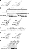
Interaction with JAM1, ZO-3, and Pals1. (A and B) Interaction with JAM1. (A) In vitro pulldown assay using a GST-fused C-terminal cytoplasmic domain of JAM1 (JAM1 C-term) or the cytoplasmic domain of JAM1 lacking the PDZ-binding motif, SFLV (JAM1 C-termΔSFLV), and lysates of cells expressing MUPP1-myc or Patj-myc. Bound proteins were eluted and analyzed by immunoblotting with anti-myc PAb. MUPP1 and Patj bound to JAM1 C-term, but not to JAM1 C-termΔSFLV. (B) In vitro pulldown assay using GST-JAM1 C-term and lysates of E. coli expressing MBP-fused PDZ domains of MUPP1 and Patj. The PDZ3 domain of MUPP1 and the PDZ3 domain of Patj specifically interacted with JAM1. (C and D) Interaction with ZO-3. (C) In vitro pulldown assay using a GST-fused C terminus of ZO-3 (ZO-3 C-term) and the C terminus of ZO-3 lacking the PDZ-binding motif, ATDL (ZO-3 C-termΔATDL), and lysates of cells expressing MUPP1-myc or Patj-myc. MUPP1 and Patj bound to ZO-3 C-term but not to ZO-3 C-termΔATDL. (D) β-Galactosidase assay using ZO-3 C-term (cloned in pGAD424 HA) and the PDZ domains of MUPP1 and Patj (in pBTM116). The PDZ7 domain of MUPP1 and the PDZ6 domain of Patj showed specific interaction with ZO-3. (E and F) Interaction with Pals1. (E) In vitro pulldown assay using GST-fused aa 1 to 122, aa 1 to 185, and aa 1 to 264 of Pals1 and lysates of cells expressing MUPP1-myc or Patj-myc. MUPP1 and Patj bound to aa 1 to 185 (containing L27N domain) and aa 1 to 264 (L27N and L27C domains), but not to aa 1 to 122 (no particular domains), of Pals1. Signals for Pals1-bound MUPP1 were weaker than those for Patj. (F) In vitro pulldown assay using GST-Pals1 constructs and the MBP-fused L27 domain of MUPP1 and Patj. MUPP1-L27 and Patj-L27 interacted with aa 1 to 185 and aa 1 to 264, but not with aa 1 to 122, of Pals1. Asterisks indicate bound MBP-fused proteins. CBB, Coomassie brilliant blue.
FIG. 3.
FIG. 3.
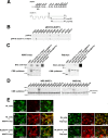
Interaction with nectin-1α and -2α. (A) Yeast two-hybrid screening for MUPP1-binding proteins with a mouse embryonic cDNA library. The PDZ2 domain of MUPP1 was used as bait, and 30 positive clones were obtained. Two of these clones encoded the C-terminal cytoplasmic domain of nectin-1α (prey no. 99 and no. 456, encoding aa 381 to 515 and aa 389 to 515, respectively). TM, transmembrane domain. (B) β-Galactosidase assay using filters and the PDZ domains of MUPP1 (in pBTM116) and the cytoplasmic domain of nectin-1α (in pVP16; prey no. 99). The PDZ2 domain of MUPP1 showed specific interaction with nectin-1α. (C) In vitro pulldown assays using a GST-fused C terminus of nectin-1α or -2α and PDZ-binding motif-deleted mutants (nectin-1α C-termΔEWYV and nectin-2α C-termΔAVYV) and lysates of cells expressing MUPP1-myc or Patj-myc. MUPP1 and Patj bound to nectin-1α and -2α but not to their PDZ-binding motif-deleted mutants. The affinity of Patj for nectins was lower than that of MUPP1. CBB, Coomassie brilliant blue. (D) In vitro pulldown assay using GST-fused nectin-2α C-term and MBP-fused PDZ domains of MUPP1 and Patj. The PDZ5 domain of MUPP1 and the PDZ5 domain of Patj specifically interacted with nectin-2α. (E) MUPP1-myc (b and c) or Patj-myc (d and e) was stably expressed in L cells that stably express nectin-2α (NL cells) (a, b, and d) or nectin-2αΔAVYV (NLΔAVYV cells) (c and e). Cells were then stained with anti-myc PAb and anti-nectin-2 MAb. As previously reported (48, 71), both nectin-2α and nectin-2αΔAVYV were concentrated at cell-cell contact sites (arrowheads). Nectin-2α, but not nectin-2αΔAVYV, recruited MUPP1 and Patj to these cell-cell contact sites. Bar, 10 μm.
FIG. 4.
FIG. 4.
Involvement of nectins in the junctional recruitment of MUPP1. (A) EpH4 cells were stably transfected with MUPP1-myc (a), MUPP1ΔPDZ2,5-myc (b), or MUPP1ΔPDZ2,3,4,5-myc (c) at comparable expression levels (data not shown) and were grown to confluence on glass coverslips. Staining with anti-myc PAb revealed that MUPP1-myc clearly localized to TJs, whereas the localization of MUPP1ΔPDZ2,5-myc and MUPP1ΔPDZ2,3,4,5-myc to TJs was slightly blurred. (B) Confluent cultures were scratched with a needle, and after culturing for 6 h, they were fixed and stained with anti-nectin-2 MAb and anti-myc PAb. MUPP1-myc accumulated at most nectin-2-positive cell-cell junctions (a [arrowheads]), whereas MUPP1ΔPDZ2,5-myc (b) and MUPP1ΔPDZ2,3,4,5-myc (c) did not (arrows). (C) Quantification of the results shown in panel B. Percentages of nectin-2-positive cell-cell junctions where the respective MUPP1 constructs were colocalized at 6 h after wounding are shown (n = 3).
FIG. 5.
FIG. 5.
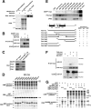
Interaction with Par6. (A) In vitro pulldown assay using GST-Par6 (Par6A) and lysates of cells expressing MUPP1-myc or Patj-myc. Both MUPP1 and Patj interacted with Par6, but the interaction of MUPP1 was weaker than that of Patj. CBB, Coomassie brilliant blue. (B) Coimmunoprecipitation assay. MUPP1-myc or Patj-myc was coexpressed in HEK293 cells with HA-Par6 (Par6B). Cells were lysed and immunoprecipitated (IP) with anti-myc PAb, followed by immunoblotting with anti-HA MAb and anti-myc PAb. HA-Par6 was present in both MUPP1-myc and Patj-myc immunoprecipitates but was more abundant in the latter. (C) Immunoprecipitation of endogenous MUPP1 from EpH4 cells. Par6 (Par6B) and Pals1, but not Patj, coimmunoprecipitated with MUPP1. (D) In vitro pulldown assay using GST-Par6 (Par6A) and MBP-fused PDZ domains of Patj. GST-Par6 specifically interacted with MBP-PDZ4. GST did not interact with any of PDZ domains. (E) In vitro pulldown assays using several GST-fused deletion constructs of Par6 (Par6A) and lysates of cells exogenously expressing MUPP1-myc, Patj-myc, or nothing. Eluates were immunoblotted with anti-myc PAb and anti-aPKC PAb. The domain between PB1 and semi-CRIB of Par6 was involved in the interaction with MUPP1 and Patj. On the other hand, PB1 alone was necessary and sufficient for the interaction with aPKC. (F) Patj-myc was coexpressed in HEK293 cells with HA-Par6 (Par6B) and/or HA-PKCζ, as indicated, followed by immunoprecipitation with anti-myc PAb. PKCζ did not significantly coprecipitate with Patj but instead inhibited the interaction of Patj with Par6. (G) In vitro pulldown assays using different recombinant proteins. The His-PKCζ RD (containing the Par6-binding region), as well as MBP-Patj PDZ4, interacted with GST-Par6 (Par6A) but not with GST. Addition of increasing amounts of MBP-Patj PDZ4 to the reaction mixture accordingly inhibited the binding of His-PKCζ RD to GST-Par6. In a control experiment, MBP-Patj PDZ1 did not bind to GST-Par6 and had no effect on the Par6-PKCζ interaction. Note that in the coimmunoprecipitation assays whose results are presented in panels B and F, the Par6B isoform but not the Par6A isoform was used, but those two isoforms exhibit similar binding profiles with respect to MUPP1 and Patj (see Fig. S3 in the supplemental material).
FIG. 6.
FIG. 6.
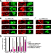
Effects of expression of MUPP1 or Patj or their L27 domains. (A) EpH4 cells were transfected with MUPP1-myc (a, b, and c) or Patj-myc (d, e, and f) and stained with anti-MUPP1 MAb (a and d), anti-Patj PAb (b and e), or anti-Pals1 PAb (c and f). MUPP1-myc as well as Patj-myc localized to TJs, with a concomitant appearance at apical membranes and in the cytoplasm. (b) At TJs between cells expressing MUPP1-myc, localization of endogenous Patj to TJs was inhibited. (d) On the other hand, expression of Patj-myc inhibited the localization of endogenous MUPP1. Localization of Pals1 was evidently inhibited by expression of MUPP1-myc (c) but not by expression of Patj-myc (f). (B) EpH4 cells were transfected with myc-MUPP1-L27 (a, b, and c) or myc-Patj-L27 (d, e, and f) and stained as described for panel A. Both myc-MUPP1-L27 and myc-Patj-L27 distributed mostly in the cytoplasm and similarly abrogated the localization of endogenous MUPP1, Patj, and Pals1 to TJs. (C) EpH4 cells were transfected with myc-MUPP1-L27 and stained with anti-Par6 PAb (a), anti-aPKC PAb (b), or anti-Par3 PAb (c). Localization of Par6 and aPKC was severely impaired, whereas that of Par3 was mostly normal at TJs between cells expressing myc-MUPP1-L27. In panels A to C, asterisks indicate cells expressing the myc constructs, and arrowheads point to the locations of cell-cell junctions between them. Bars, 10 μm. (D) Quantification of the results shown in panels A to C. Percentages of cell-cell junctions where junctional proteins were appropriately targeted were calculated for each set of conditions. Note that the localization of endogenous MUPP1 and Patj could not be examined when full-length MUPP1-myc and Patj-myc were expressed, respectively (asterisks). A total of 52 to 149 cell-cell junctions were examined for each set of conditions. GFP, green fluorescent protein.
FIG. 7.
FIG. 7.
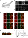
Knockdown of MUPP1 and Patj by RNAi. (A) Immunoblotting with anti-MUPP1 MAb and anti-Patj PAb of lysates of control, two MUPP1 RNAi (MUPP1 KD-1 and MUPP1 KD-2), two Patj RNAi (Patj KD-1 and Patj KD-2), and two MUPP1 and Patj RNAi (MUPP1 KD-2 Patj KD-1 and MUPP1 KD-2 Patj KD-2) stable EpH4 cell lines. Blotting for γ-tubulin was used as a loading control. (B) Control, MUPP1 KD-2, Patj KD-2, and MUPP1 KD-2 Patj KD-1 cells were grown to confluence on glass coverslips and stained with anti-MUPP1 MAb, anti-Patj PAb, or anti-ZO-1 MAb. TJs were linearly established in control and MUPP1 KD-2 cells, whereas they were fragmentary in Patj KD-2 cells and MUPP1 KD-2 Patj KD-1 cells. Bar, 10 μm. (C) MUPP1 KD-2 and Patj KD-2 cells were stained with anti-Pals1 PAb (a and f), anti-Par6 PAb (b and g), anti-aPKC PAb (c and h), anti-Par3 PAb (d and i), and anti-E-cadherin MAb (e and j). In MUPP1 KD-2 cells, the localization of these junctional proteins was normal (a to e). In Patj KD cells, Pals1, Par6, and aPKC were severely delocalized from ZO-1-positive fragmental junctions, whereas Par3 and E-cadherin mostly colocalized with ZO-1 (arrowheads). Bar, 10 μm. (D) MUPP1 KD-2 and Patj KD-2 cells were grown on Transwell filters and stained with anti-ZO-1 MAb (green) and anti-Syntaxin3 PAb (red) (upper panels) and with anti-ZO-1 MAb (green) and anti-ErbB2 PAb (red) (lower panels). Vertical sectional images were generated by confocal microscopy. In MUPP1 KD-2 cells, Syntaxin3 and ErbB2 localized to apical membranes and lateral membranes, respectively, whereas in Patj KD-2 cells, they were localized to both apical and lateral membranes. Bar, 10 μm. (E) TER of control, MUPP1 KD-1, MUPP1 KD-2, Patj KD-1, Patj KD-2, MUPP1 KD-2 Patj KD-1, and MUPP1 KD-2 Patj KD-2 cells. The knockdown of Patj significantly affected the development of the epithelial barrier (n = 4 for each cell line).
FIG. 8.
FIG. 8.
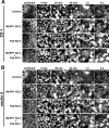
Ca2+-switch assay. Control, MUPP1 KD-2, Patj KD-2, and MUPP1 KD-2 Patj KD-1 cells were grown to confluence on glass coverslips. They were then transferred to a low-Ca2+ medium overnight to disassemble the junctions and returned to normal culture medium. At different time points after the addition of the medium, cells were fixed and stained with anti-ZO-1 PAb (A) and anti-nectin-2 MAb (B). Formation of ZO-1-positive TJs as well as nectin-2-positive AJs was severely retarded by the knockdown of Patj. Bar, 10 μm.
FIG. 9.
FIG. 9.
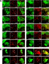
Attempts to rescue aberrant TJs in Patj KD cells. (A) MUPP1 KD-2+Patj KD-1 cells were stably transfected with MUPP1-myc (a and g), Patj-myc (b and h), PatjΔL27-myc (c and i), PatjΔ4-myc (d and j), PatjΔ2,5-myc (e and k), and myc-Patj L27 (f and l) and stained with anti-ZO-1 PAb (a to f) or anti-Pals1 PAb (g to l). The expression levels of the constructs were similar (data not shown). Expression of Patj, PatjΔ4, and PatjΔ2,5 restored the development of linear TJs (b, d, and e [arrowheads]), whereas that of MUPP1, PatjΔL27, and Patj L27 was ineffective (a, c, and f). Expression of Patj, PatjΔ4, and PatjΔ2,5 also restored the junctional localization of Pals1 (h, j, and k), but that of MUPP1, PatjΔL27, and Patj L27 did not (g, i, and l). Note that except for Patj-L27, which does not cover the targeted region of RNAi, point mutations were introduced into all Patj constructs to attain RNAi resistance. Asterisks indicate cells expressing the myc constructs. Bars, 10 μm. (B) Patj KD-1 cells were transfected with myc-Cdc42G12V (a and c) or myc-Rac1G12V (b and d) and stained with anti-ZO-1 MAb (a and b) or anti-Pals1 PAb (c and d). Expression of Cdc42G12V restored the junctional accumulation of ZO-1 and Pals1 in a linear fashion (a and c [arrowheads]). By contrast, Rac1G12V was ineffective (b and d). Asterisks indicate cells expressing the myc constructs. Bars, 10 μm.

Similar articles

Cited by

References

    1. Ackermann, F., N. Zitranski, D. Heydecke, B. Wilhelm, T. Gudermann, and I. Boekhoff. 2008. The Multi-PDZ domain protein MUPP1 as a lipid raft-associated scaffolding protein controlling the acrosome reaction in mammalian spermatozoa. J. Cell. Physiol. 214757-768. - PubMed
    1. Adachi, M., A. Inoko, M. Hata, K. Furuse, K. Umeda, M. Itoh, and S. Tsukita. 2006. Normal establishment of epithelial tight junctions in mice and cultured cells lacking expression of ZO-3, a tight junction MAGUK protein. Mol. Cell. Biol. 269003-9015. - PMC - PubMed
    1. Anderson, J. M., and C. M. Van Itallie. 1995. Tight junctions and the molecular basis for regulation of paracellular permeability. Am. J. Physiol. 269G467-G475. - PubMed
    1. Ando-Akatsuka, Y., S. Yonemura, M. Itoh, M. Furuse, and S. Tsukita. 1999. Differential behavior of E-cadherin and occludin in their colocalization with ZO-1 during the establishment of epithelial cell polarity. J. Cell. Physiol. 179115-125. - PubMed
    1. Aoki, J., S. Koike, H. Asou, I. Ise, H. Suwa, T. Tanaka, M. Miyasaka, and A. Nomoto. 1997. Mouse homolog of poliovirus receptor-related gene 2 product, mPRR2, mediates homophilic cell aggregation. Exp. Cell Res. 235374-384. - PubMed

Publication types

MeSH terms

Substances

LinkOut - more resources