. 2009 Aug;50(8):3985-95.
doi: 10.1167/iovs.08-3364. Epub 2009 Feb 28.

Age-dependent disease expression determines remodeling of the retinal mosaic in carriers of RPGR exon ORF15 mutations

Affiliations

Age-dependent disease expression determines remodeling of the retinal mosaic in carriers of RPGR exon ORF15 mutations

William A Beltran et al. Invest Ophthalmol Vis Sci. 2009 Aug.

Abstract

Purpose: To characterize the retinal histopathology in carriers of X-linked progressive retinal atrophy (XLPRA1 and XLPRA2), two canine models of X-linked retinitis pigmentosa caused, respectively, by a stop and a frameshift mutation in RPGRORF15.

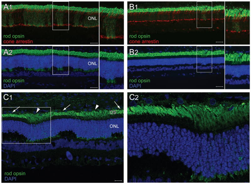
Methods: Retinas of XLPRA2 and XLPRA1 carriers of different ages were processed for morphologic evaluation, TUNEL assay, and immunohistochemistry. Cell-specific markers were used to examine retinal remodeling events.

Results: A mosaic pattern composed of patches of diseased and normal retina was first detected in XLPRA2 carriers at 4.9 weeks of age. A peak of photoreceptor cell death led to focal rod loss; however, in these patches an increased density of cones was found to persist over time. Patches of disease gradually disappeared so that by 39 weeks of age the overall retinal morphology, albeit thinner, had improved lamination. In older XLPRA2 carriers (>or=8.8 years), extended regions of severe degeneration occurred in the peripheral/mid-peripheral retina. In XLPRA1 carriers, opsin mislocalization and rare events of rod death were detected by TUNEL assay at 20 weeks of age; however, only patchy degeneration was seen by 1.4 years and was still apparent at 7.8 years.

Conclusions: The time of onset and the progression of the disease differed between the two models. In the early-onset form (XLPRA2) the morphologic appearance of the retinal mosaic changed as a function of age, suggesting that structural plasticity persists in the early postnatal canine retina as mutant photoreceptors die. In the late-onset form (XLPRA1), patches of disease persisted until later ages.


Figures

Figure 1
Figure 1
Retinal morphology in carriers of XLPRA2. (A) Normal maturation of the retina at 3.9 weeks. (B) Early patch of outer segment misalignment (below white bar) and ONL thickening in a 4.9 week-old carrier. (C) Prominent foci of outer segment disruption in a 5.9 week-old carrier. Note the increased thickness of the ONL, decreased cell density and occasional pyknotic figures (arrows) in these patches. (D1) Multifocal retinal degeneration in a 26.1 week-old carrier of XLPRA2. Foci of retinal degeneration (arrows) are surrounded by areas of normal retinal morphology. (D2) High magnification of a patch of retinal degeneration. Note the misalignment and loss of photoreceptor outer segments, the shortening and broadening of the inner segments, and the decrease in cell density in the ONL. (D3) High magnification at the transition zone of a patch of retinal disease. Note the progressive disorganization of the inner and outer segments, and the decrease in chromatin compaction of the rods as one moves from a normal to a diseased area. (E1–3) Retinal disease in a 10.3 year-old carrier of XLPRA2. (E1) Despite a reduction in ONL thickness (approx 5–6 nuclei thick) in the central region, the retinal morphology appears well preserved in comparison to the severe retinal degeneration and complete retinal atrophy observed, respectively, in the mid-peripheral (E2) and peripheral (E3) regions. (Epoxy resin, 1 μm sections) Scale bars: (A–C, E1–3) 20 μm; (D1) 80 μm. Abbreviations: ONL = outer nuclear layer; INL = inner nuclear layer; GCL = ganglion cell layer.
Figure 2
Figure 2
Retinal morphology in carriers of XLPRA1. (A1–3) H&E stained retinal cryosections along the superior meridian of a 24 week-old carrier of XLPRA1. No morphologic abnormalities are detected at this age in the central (A1), mid-peripheral (A2), and peripheral (A3) regions. (B1–3) Epoxy resin retinal section of a 1.4 year-old carrier of XLPRA1. (B1) Focal patches (arrows) of retinal degeneration are found throughout the retina. In these areas, there is a loss of OS, shortening and broadening of IS and a decrease in ONL cell density (B3) and contrast with surrounding areas (B2) that have a better preserved morphology. (C1–3) Epoxy resin retinal section of a 7.9 year-old carrier of XLPRA1. (C1) A generalized thinning of the ONL is seen throughout the retina, yet remaining photoreceptors maintain their normal IS and OS (asterisks). (C2) Focal area of severe ONL thinning. (C3) Higher magnification of C2 insert. In this foci note the increased number of cone IS and nuclei, and the reduced density of rods. Scale bars: (B2–3) 10 μm; (A1–3, C1–2) 20 μm; (B1, C3) 40 μm. Abbreviations: OS = outer segments; IS = inner segments; ONL = outer nuclear layer; INL = inner nuclear layer; GCL = ganglion cell layer.
Figure 3
Figure 3
Disease and remodeling of the rod photoreceptor mosaic in carriers of XLPRA2. (A1–2) Double fluorescence immunohistochemistry in a 4.9 week-old XLPRA2 carrier shows numerous patches of rod opsin (green) mislocalization to the inner segments and ONL but no abnormalities in cone arrestin labeling (red) (A1). Co-labeling with DAPI nuclear stain (blue) shows that patches of rod opsin mislocalization and rod neurite sprouting (see arrow in insert) occur in areas of ONL thickening (A2). Double fluorescence immunohistochemistry in a 39 week-old XLPRA2 carrier shows a single patch of rod opsin (green) mislocalization to the inner segments and persistent normal expression of cone arrestin (B1). DAPI nuclear stain shows that the overall ONL thickness is reduced (B2). The inner margin of the ONL is less wavy than at earlier ages. However persistent focal regions of ONL thickening are seen at the sites of rod opsin mislocalization (B2). Rod opsin and DAPI labeling in a 5.9 week-old XLPRA2 carrier imaged by confocal microscopy (maximal projection reconstituted image) (C1). Note the disorganized OS (arrowheads) in the patches of opsin mislocalization and ONL thickening, which contrast with the aligned rod OS (arrows) in adjacent areas of non-diseased retina. Insert seen in C1 is shown as a single plane image at higher magnification in (C2). Scale bars A1–2, B1–2: 40 μm; C1: 20 μm. Abbreviations: ONL = outer nuclear layer; OS = outer segments.
Figure 4
Figure 4
Disease and remodeling of the cone photoreceptor mosaic in carriers of XLPRA2. Double fluorescence immunohistochemistry in a 4.9 week-old XLPRA2 carrier shows M/L opsin mislocalization to the inner segments, ONL and cone pedicle (A1) in the same patches where rod opsin is mislocalized (see asterisks) (A2). (Note 1: the labeling of the inner retina with the M/L opsin antibody is not specific. Note 2: the yellow labeling seen in the cone outer segments is the result of a post-acquisition increase in the green signal of the overlay image to improve visualization of the rod opsin mislocalization, and does not represent co-localization). In a 5.9 week-old XLPRA2 carrier, faint S opsin mislocalization to the inner segments, and ONL is seen in some cones (arrows) (B1). These cones are located within patches of rod opsin mislocalization (B2). Immunolabeling in a 5.9 week-old XLPRA2 carrier show an increased density in M/L cones in the patches of rod and M/L opsin mislocalization (C1–3). (D1) Plastic embedded retinal section of a 26.1 week-old XLPRA2 carrier showing focal accumulation of large euchromatic nuclei resembling that of cones in an area of rod loss. (D2–3) Double fluorescence IHC in a 39 week-old XLPRA2 carrier showing increased density of cones that can either be distributed in a monolayer (D2) or clumped together (D3). Note that there is no rod opsin mislocalization in the patches of increased cone density. Scale bars : (D1) 10 μm; (A1–2, B1–2, C2–3, D2–3) 20 μm; (C1) 40 μm. Abbreviations: ONL = outer nuclear layer.
Figure 5
Figure 5
Focal invasion of the subretinal layer by phagocytic cells in carriers of XLPRA2 (A) Plastic embedded retinal section of a 26.1 week-old XLPRA2 dog showing a phagocytic cell in the subretinal space overlying a patch of degeneration. Note the multiple cytoplasmic inclusions suggestive of phagocytized rod disc material. (B) Double fluorescent IHC in this same animal labels shows that these cells are labeled with CD18 but not with RPE65 antibodies. This confirms their macrophage origin. Scale bar: 10 μm. Abbreviations: ONL = outer nuclear layer.
Figure 6
Figure 6
Disease and remodeling of the inner retinal mosaic in carriers of XLPRA2 at 7.9 and 39 weeks of age. Double fluorescence IHC in a 7.9 week-old XLPRA2 carrier shows focal dendritic retraction of rod bipolar cells (yellow) beneath the patches of rod opsin mislocalization (asterisks) (A1). Absence of rod bipolar dendritic retraction is noted in the 39 week-old XLPRA2 carrier (A2). (B1–2) GFAP immunohistochemistry in a 7.9 week-old XLPRA2 carrier shows patchy labeling of the radial extensions of reactive Müller cells (B1). Double IHC confirms that these GFAP labeled radial extensions occur in Müller cells located beneath the larger patches of rod opsin mislocalization (B2). Scale bars: (A1–2) 20 μm; (B1–2) 40 μm. Abbreviations: ONL = outer nuclear layer; INL = inner nuclear layer.
Figure 7
Figure 7
Photoreceptor mosaic in the XLPRA1 carrier. (A1–2) Double fluorescence immunohistochemistry in a 24 week-old XLPRA1 carrier shows numerous patches of rod opsin (green) mislocalization to the inner segments and ONL (asterisks), but no abnormalities in cone arrestin labeling (red) (A1). Co-labeling with DAPI nuclear stain (blue) shows patches of rod opsin mislocalization and rod neurite sprouting (see arrows) (A2).(B1–2) Double fluorescence immunohistochemistry in a 24 week-old XLPRA1 carrier shows M/L opsin mislocalization to the inner segments, ONL and cone pedicle (B1) in the same patches where rod opsin is mislocalized (B2). Note that M/L opsin labeling of the inner nuclear layer and ganglion cell layer is non-specific. Scale bars: (A1–2) 20 μm; (B1–2) 40 μm. Abbreviations: ONL = outer nuclear layer.
Figure 8
Figure 8
Photoreceptor cell death in carriers of XLPRA. (A) Kinetics of photoreceptor cell death in the superior meridian of XLPRA2 carriers. The insert shows previously published (Beltran et al, IOVS, 2006) kinetics of cell death in affected XLPRA2 dogs. (B) TUNEL labeling of rod nuclei (arrows) in a 4.9 week-old XLPRA2 carrier is seen exclusively in the patches of rod opsin mislocalization. (C) Single TUNEL-labeled rod in a 24 week-old XLPRA1 carrier is located in a patch of rod opsin mislocalization. Abbreviations: ONL = outer nuclear layer.
Figure 9
Figure 9
Remodeling of the retinal mosaic in carriers of XLPRA2. (A1) Numerous foci of retinal disease (horizontal black bars) occupy a significant proportion of the retinal length as seen here in the mid-peripheral region of the superior retina of a 5.9 week-old carrier of XLPRA2. (A2) Infrequent and small foci of retinal disease are found at later ages as illustrated here in the mid-peripheral region of the superior retina of a 39 week-old carrier of XLPRA2. (B) Percentage (mean ± SD) of the total retinal length along the superior and inferior meridians that is occupied by foci of retinal disease in carriers of XLPRA2 of different ages. (C) Mean (±SD) ONL thickness in the mid-peripheral region of the superior and inferior meridians in XLPRA2 carriers of different ages. Note that ONL thickness was measured in areas between patches of disease. Scale bars: 10 μm.

Similar articles

Cited by

References

    1. Bird AC. X-linked retinitis pigmentosa. Br J Ophthalmol. 1975;59:177–199. - PMC - PubMed
    1. Jay M. On the heredity of retinitis pigmentosa. Br J Ophthalmol. 1982;66:405–416. - PMC - PubMed
    1. Vervoort R, Lennon A, Bird AC, et al. Mutational hot spot within a new RPGR exon in X-linked retinitis pigmentosa. Nat Genet. 2000;25:462–466. - PubMed
    1. Bader I, Brandau O, Achatz H, et al. X-linked retinitis pigmentosa: RPGR mutations in most families with definite X linkage and clustering of mutations in a short sequence stretch of exon ORF15. Invest Ophthalmol Vis Sci. 2003;44:1458–1463. - PubMed
    1. Sharon D, Sandberg MA, Rabe VW, Stillberger M, Dryja TP, Berson EL. RP2 and RPGR mutations and clinical correlations in patients with X-linked retinitis pigmentosa. Am J Hum Genet. 2003;73:1131–1146. - PMC - PubMed

Publication types

MeSH terms

Substances