. 2009 Mar 1;91(3):652-63.
doi: 10.2106/JBJS.H.01177.

Identification of progenitor cells that contribute to heterotopic skeletogenesis

Affiliations

Identification of progenitor cells that contribute to heterotopic skeletogenesis

Vitali Y Lounev et al. J Bone Joint Surg Am. .

Abstract

Background: Individuals who have fibrodysplasia ossificans progressiva develop an ectopic skeleton because of genetic dysregulation of bone morphogenetic protein (BMP) signaling in the presence of inflammatory triggers. The identity of progenitor cells that contribute to various stages of BMP-induced heterotopic ossification relevant to fibrodysplasia ossificans progressiva and related disorders is unknown. An understanding of the cellular basis of heterotopic ossification will aid in the development of targeted, cell-specific therapies for the treatment and prevention of heterotopic ossification.

Methods: We used Cre/loxP lineage tracing methods in the mouse to identify cell lineages that contribute to all stages of heterotopic ossification. Specific cell populations were permanently labeled by crossing lineage-specific Cre mice with the Cre-dependent reporter mice R26R and R26R-EYFP. Two mouse models were used to induce heterotopic ossification: (1) intramuscular injection of BMP2/Matrigel and (2) cardiotoxin-induced skeletal muscle injury in transgenic mice that misexpress BMP4 at the neuromuscular junction. The contribution of labeled cells to fibroproliferative lesions, cartilage, and bone was evaluated histologically by light and fluorescence microscopy. The cell types evaluated as possible progenitors included skeletal muscle stem cells (MyoD-Cre), endothelium and endothelial precursors (Tie2-Cre), and vascular smooth muscle (Smooth Muscle Myosin Heavy Chain-Cre [SMMHC-Cre]).

Results: Vascular smooth muscle cells did not contribute to any stage of heterotopic ossification in either mouse model. Despite the osteogenic response of cultured skeletal myoblasts to BMPs, skeletal muscle precursors in vivo contributed minimally to heterotopic ossification (<5%), and this contribution was not increased by cardiotoxin injection, which induces muscle regeneration and mobilizes muscle stem cells. In contrast, cells that expressed the vascular endothelial marker Tie2/Tek at some time in their developmental history contributed robustly to the fibroproliferative, chondrogenic, and osteogenic stages of the evolving heterotopic endochondral anlagen. Importantly, endothelial markers were expressed by cells at all stages of heterotopic ossification. Finally, muscle injury and associated inflammation were sufficient to trigger fibrodysplasia ossificans progressiva-like heterotopic ossification in a setting of chronically stimulated BMP activity.

Conclusions: Tie2-expressing progenitor cells, which are endothelial precursors, respond to an inflammatory trigger, differentiate through an endochondral pathway, contribute to every stage of the heterotopic endochondral anlagen, and form heterotopic bone in response to overactive BMP signaling in animal models of fibrodysplasia ossificans progressiva. Thus, the ectopic skeleton is not only supplied by a rich vasculature, but appears to be constructed in part by cells of vascular origin. Further, these data strongly suggest that dysregulation of the BMP signaling pathway and an inflammatory microenvironment are both required for the formation of fibrodysplasia ossificans progressiva-like lesions.


Figures

Fig. 1
Fig. 1
Schematic of the Cre/loxP lineage tracing methodology. Transgenic mice expressing the Cre recombinase under the control of a cell-specific promoter (A) are crossed to reporter mice (B) in which a reporter gene (e.g., lacZ in R26R mice) is separated from a constitutively active promoter by transcriptional stop sequences that are flanked by loxP sites, the target sequences recognized by Cre recombinase. Only cells of cell-specific Cre X loxP-lacZ offspring that express Cre undergo DNA excision of the stop sequences, resulting in transcription of the reporter gene (C). Note that loxP recombination at any point in a cell's developmental history results in permanent labeling of that cell and its daughters, regardless of whether Cre expression persists. In our experiments, the Cre-dependent reporters R26R and R26R-EYFP were used.
Fig. 2
Fig. 2
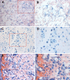
Contribution of MyoD+ and Tie2+ cells to heterotopic ossification following intramuscular injection of BMP2. A, B, and C: MyoDiCre;R26R-EYFP mice. D through I: Tie2-Cre;R26R mice. A: Phase image showing robust fibroproliferative response (FP) adjacent to skeletal muscle fibers (M), shown in transverse section, eight days after injection of BMP2. B: Fluorescence image of section in A. Only a few EYFP-labeled cells (examples at arrowheads) are present in the fibroproliferative lesion, whereas essentially all muscle fibers (M) are labeled. C: DAPI image showing the density of fibroproliferative cells. D: Longitudinal section of control, uninjected, muscle stained with X-gal. Tie2-Cre;R26R mice exhibit extensive β-gal labeling of the vasculature (blue staining). E: The majority of fibroproliferative cells (FP) four days after injection are intensely stained for β-gal. Longitudinal profiles of muscle fibers (M) are shown. F: At seven days after injection, the preosseous anlagen is composed of both fibroproliferative tissue (FP) and chondrogenic cells (C). In this section, representation of β-gal-labeled cells in the cartilage anlagen is lower than average. G and H: Typical examples of cartilage labeling from two additional mice, seven days after injection. Substantial regional variation in the extent of labeling is sometimes observed (compare left and right regions of H). I: New lamellar bone (B) fourteen days after BMP2 injection, showing β-gal-labeled osteocytes (two shown with arrowheads). Marrow elements (m) are intensely stained because of Tie2-Cre expression in hematopoietic lineages (original magnification, ×200).
Fig. 3
Fig. 3
Cardiotoxin injury of skeletal muscle stimulates and synchronizes heterotopic ossification in Nse-BMP4 transgenic mice. A and B: Radiographs of wild-type (A) and Nse-BMP4 (B) mice made three weeks after cardiotoxin injection. The white arrow indicates heterotopic ossification, which was observed only in the Nse-BMP4 transgenic mouse. Control injections with phosphate-buffered saline solution did not result in heterotopic ossification. C through F: Histological sections of lesions from cardiotoxin-injected Nse-BMP4 mice at one, four, seven, and fourteen days after injection, showing the morphology and features of heterotopic ossification. C: Muscle degeneration and lymphocytic infiltration (small round cells) is apparent at day 1. Two muscle fibers (M) are marked. D: A robust fibroproliferative response is apparent by four days. Fibroproliferative tissue (FP) is interspersed among the remaining muscle fibers (M). E: Section showing extensive cartilage differentiation (C) at seven days. Fibroproliferative cells (FP) are also visible in this field. F: At fourteen days, heterotopic bone (B; light blue) with marrow elements (m; dark blue) is present. (Safranin O staining; cartilage matrix is orange to red, and nuclei are blue; original magnification, ×200).
Fig. 4
Fig. 4
Tie2+ cells contribute to all stages of heterotopic ossification after cardiotoxin-induced muscle injury in Tie2-Cre;R26R;Nse-BMP4 transgenic mice. A and B: Section of fibroproliferative tissue four days after cardiotoxin injection into the quadriceps muscle. X-gal staining revealed many β-gal-positive cells (blue). Labeled cells include both rounded lymphocyte-like cells and fibroblastic cells. Panel B represents a higher magnification of the boxed region in A. C and D: Low and high-power magnification images of chondrogenic area developing in muscle seven days after cardiotoxin injection. Approximately 50% of cartilage cells are X-gal stained (blue). E and F: Low and high-power magnification images of developing bone fourteen days after cardiotoxin injection. The majority of osteocytes were positive for β-gal. All sections were counterstained with eosin (original magnification, ×100 for A, C, and E and ×400 for B, D, and F).
Fig. 5
Fig. 5
Endothelial markers are expressed at all stages of the endochondral anlagen in BMP4-associated heterotopic ossification. Stages of endochondral ossification: fibroproliferative (A, D, G, and J), chondrogenic (B, E, H, and K), and osteogenic (C, F, I, and L). Sections were indirectly labeled for CD144 (A, B, and C), von Willebrand Factor (D, E, and F), and Tie2 (G, H, and I). Control sections in which nonspecific serum replaced the primary antibody (J, K, and L) showed no immunoreactivity. Nuclei (blue) were stained with DAPI, and immunostaining and DAPI images were merged. Each stage is a mixture of immunoreactive and nonreactive cells, consistent with the lineage tracing results (Figs. 2 and 4) (original magnification, ×600).
Fig. 6
Fig. 6
Working model of BMP-associated heterotopic ossification. Injury to skeletal muscle and connective tissue leads to monocyte invasion, macrophage activation, tissue hypoxia, and upregulation of inflammatory cytokines and osteogenic factors including BMPs that recruit Tie2-expressing progenitor cells to form the heterotopic anlagen,,-. Wound hypoxia and inflammatory cytokines contribute to the angiogenic response in wound-healing, at least in part, by upregulating the expression of Tie2 mRNA and protein in these endothelial cells. Hypoxia-related pH changes may further sensitize fibrodysplasia ossificans progressiva cells to ambient levels of BMPs, which further upregulates Tie2 expression and subsequent endothelial cell mobilization and migration. The inflammatory reaction to muscle injury, the secretion of BMPs, and the cross-talk between cells of the innate and adaptive immune system stimulate the induction and propagation of an ectopic skeletal element. The blue lines indicate hematopoietic-derived pathways; brown lines, connective tissue progenitor-derived pathways; black lines, basal and very early post-traumatic conditions; green lines, muscle-derived pathways; blunt-end lines, inhibitory pathways; and arrows, stimulatory pathways. PC = progenitor cells, HSC = hematopoietic stem cells, T = T-cells, B = B-cells, MA = mast cells, FP = fibroproliferative cells, CB = chondroblasts, OB = osteoblasts, PGE2 = prostaglandin E2, and TGF-β = transforming growth factor-beta.

Similar articles

Cited by

References

    1. Kaplan FS, Groppe J, Pignolo RJ, Shore EM. Morphogen receptor genes and metamorphogenes: skeleton keys to metamorphosis. Ann N Y Acad Sci. 2007;1116:113-33. - PubMed
    1. Schmierer B, Hill CS. TGFbeta-SMAD signal transduction: molecular specificity and functional flexibility. Nat Rev Mol Cell Biol. 2007;8:970-82. - PubMed
    1. Shore EM, Xu M, Feldman JG, Fenstermacher DA, Choi TJ, Choi IH, Connor JM, Delai P, Glasser DL, LeMerrer M, Morhart R, Rogers JG, Smith R, Triffitt JT, Urtizberea JA, Zasloff M, Brown MA, Kaplan FS. A recurrent mutation in the BMP type I receptor ACVR1 causes inherited and sporadic fibrodysplasia ossificans progressiva. Nat Genet. 2006;38:525-7. - PubMed
    1. Wozney JM, Rosen V, Celeste AJ, Mitsock LM, Whitters MJ, Kriz RW, Hewick RM, Wang EA. Novel regulators of bone formation: molecular clones and activities. Science. 1988;242:1528-34. - PubMed
    1. Urist MR. Bone: formation by autoinduction. Science. 1965;150:893-9. - PubMed

Publication types

MeSH terms