. 2009 Mar 17;119(10):1386-97.
doi: 10.1161/CIRCULATIONAHA.108.802918. Epub 2009 Mar 2.

Divergent tumor necrosis factor receptor-related remodeling responses in heart failure: role of nuclear factor-kappaB and inflammatory activation

Affiliations

Divergent tumor necrosis factor receptor-related remodeling responses in heart failure: role of nuclear factor-kappaB and inflammatory activation

Tariq Hamid et al. Circulation. .

Abstract

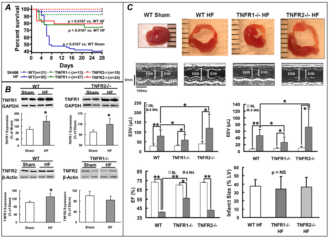
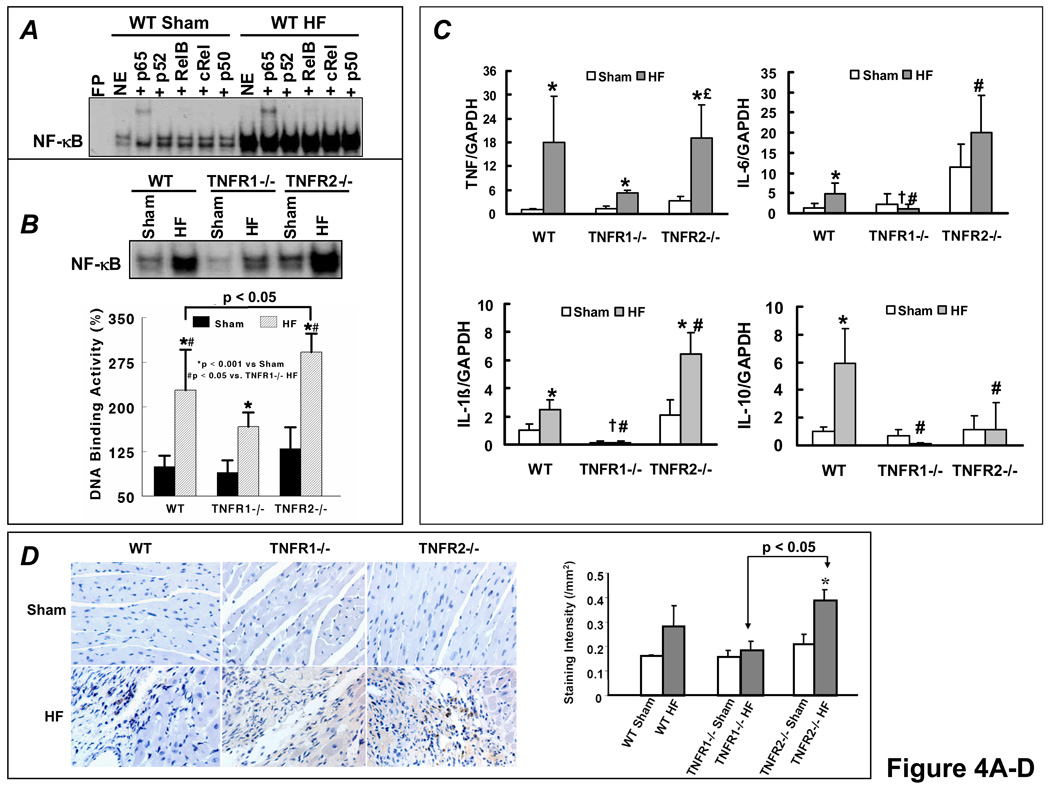
Background: Although preclinical data suggested that tumor necrosis factor-alpha (TNF) neutralization in heart failure (HF) would be beneficial, clinical trials of TNF antagonists were paradoxically negative. We hypothesized that TNF induces opposing inflammatory and remodeling responses in HF that are TNF-receptor (TNFR) specific.

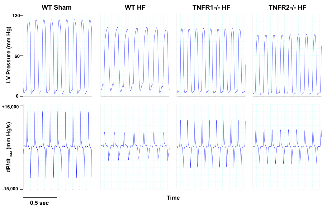
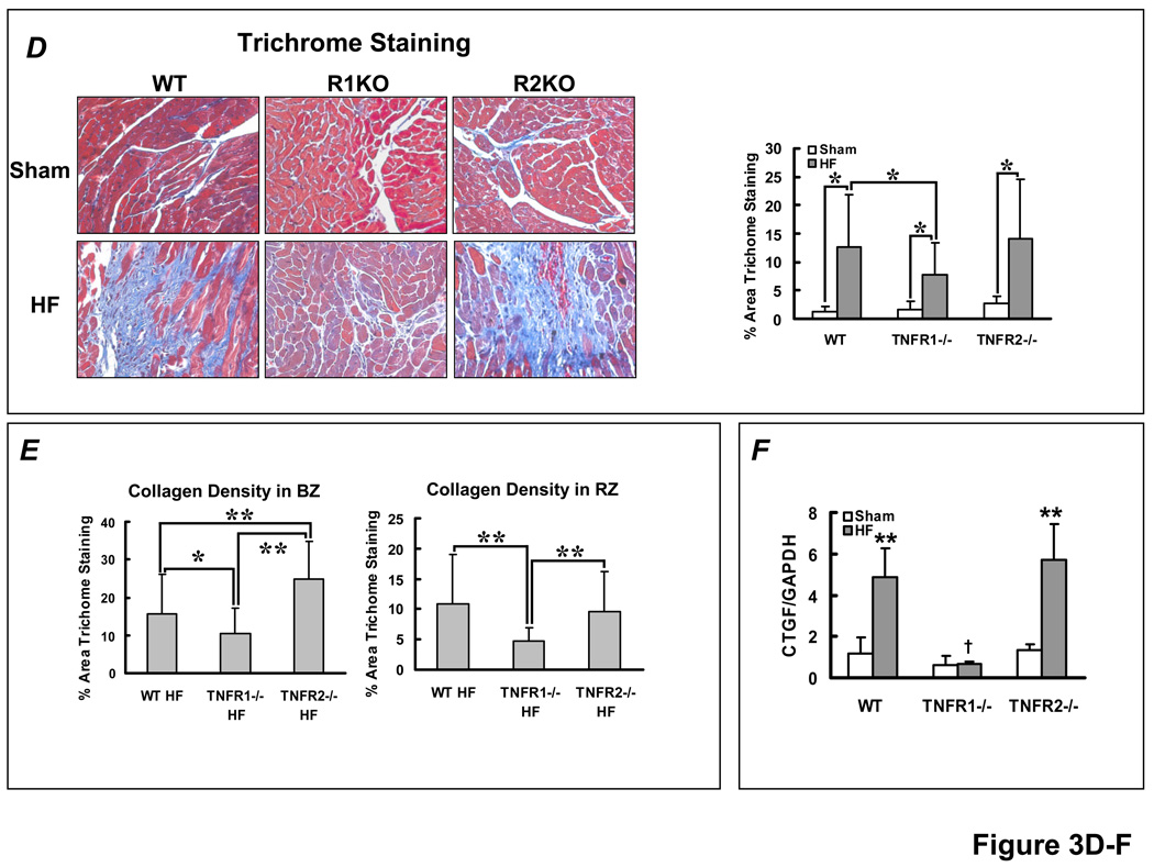
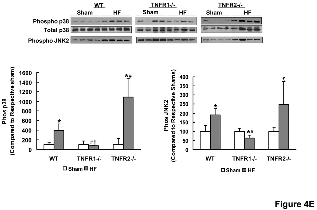
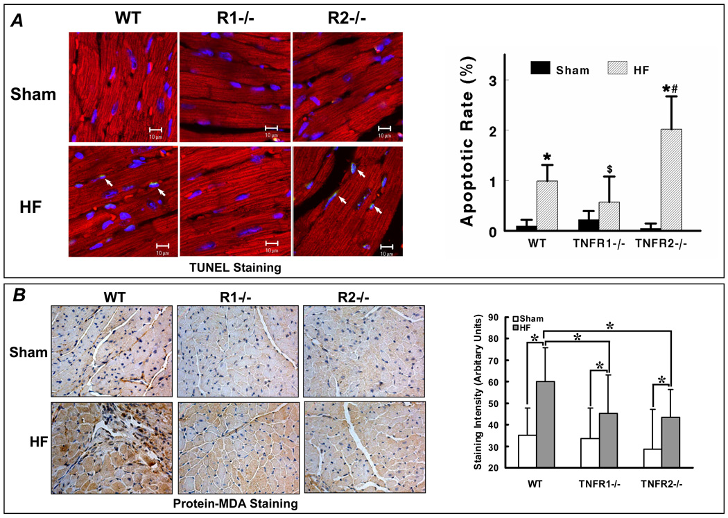
Methods and results: HF was induced in wild-type (WT), TNFR1(-/-), and TNFR2(-/-) mice via coronary ligation. Compared with WT HF, 4-week postinfarction survival was significantly improved in both TNFR1(-/-) and TNFR2(-/-) HF. Compared with sham, WT HF hearts exhibited significant remodeling with robust activation of nuclear factor (NF)-kappaB, p38 mitogen-activated protein kinase, and JNK2 and upregulation of TNF, interleukin (IL)-1beta, IL-6, and IL-10. Compared with WT HF, TNFR1(-/-) HF exhibited (1) improved remodeling, hypertrophy, and contractile function; (2) less apoptosis; and (3) diminished NF-kappaB, p38 mitogen-activated protein kinase, and JNK2 activation and cytokine expression. In contrast, TNFR2(-/-) HF showed exaggerated remodeling and hypertrophy, increased border zone fibrosis, augmented NF-kappaB and p38 mitogen-activated protein kinase activation, higher IL-1beta and IL-6 gene expression, greater activated macrophages, and greater apoptosis. Oxidative stress and diastolic function were improved in both TNFR1(-/-)and TNFR2(-/-) HF. In H9c2 cardiomyocytes, sustained NF-kappaB activation was proapoptotic, an effect dependent on TNFR1 signaling, whereas TNFR2 overexpression attenuated TNF-induced NF-kappaB activation.

Conclusions: TNFR1 and TNFR2 have disparate and opposing effects on remodeling, hypertrophy, NF-kappaB, inflammation, and apoptosis in HF: TNFR1 exacerbates, whereas TNFR2 ameliorates, these events. However, signaling through both receptors is required to induce diastolic dysfunction and oxidative stress. TNFR-specific effects in HF should be considered when therapeutic anti-TNF strategies are developed.


Conflict of interest statement

CONFLICT OF INTEREST DISCLOSURES

There are no commercial affiliations or conflicts of interest to disclose.

Figures

Figure 1
Figure 1
TNFR1 and TNFR2 differentially modulate LV remodeling. (A) Kaplan-Meier survival curves from WT, TNFR1−/−, and TNFR2−/− mice after coronary ligation (HF) or sham operation. (B) Immunoblots and corresponding group data depicting changes in TNFR1 and TNFR2 expression in failing, remodeled myocardium. *p<0.05 vs. sham. (C) Short-axis LV sections, M-mode echocardiograms, and group data for LV function and infarct size from WT, TNFR1−/−, and TNFR2−/− sham and HF mice. **p<0.005, *p<0.05.
Figure 2
Figure 2
Representative hemodynamic recordings for LV pressure and dP/dtmax from WT sham, WT HF, TNFR1−/− HF, and TNFR2−/− HF mice. LV peak pressure and dP/dtmax were depressed and LVEDP was elevated in WT HF. TNFR1−/− HF displayed global improvement in these parameters. TNFR2−/− HF exhibited similar reductions in dP/dtmax but improved LVEDP compared with WT HF.
Figure 3
Figure 3
TNFR1- and TNFR2-specific effects on hypertrophy and fibrosis in HF. (A) LV mass/tibia length ratio from WT, TNFR1−/−, and TNFR2−/− sham and HF mice. **p<0.005, *p<0.05. (B) Normalized ANF gene expression from sham and failing myocardium by qRT-PCR analysis (n=4/group). **p<0.005, *p<0.05 vs. sham, #p<0.005 vs WT and TNFR2−/− HF. (C) Representative H&E histomicrographs of remodeled myocardium from each experimental group and quantitation of myocyte cross-sectional area. **p<0.0001, *p<0.05. (D) Masson’s Trichrome stains and quantitation of fibrosis in non-infarcted myocardium (i.e., remote and border zones). *p<0.05. (E) Selective border-zone (BZ) and remote-zone (RZ) fibrosis quantitation. **p<0.001, *p<0.05. (F) Normalized CTGF gene expression by qRT-PCR, **p<0.005 vs. sham; p<0.005 vs WT and TNFR2−/− HF (n = 6/group).
Figure 3
Figure 3
TNFR1- and TNFR2-specific effects on hypertrophy and fibrosis in HF. (A) LV mass/tibia length ratio from WT, TNFR1−/−, and TNFR2−/− sham and HF mice. **p<0.005, *p<0.05. (B) Normalized ANF gene expression from sham and failing myocardium by qRT-PCR analysis (n=4/group). **p<0.005, *p<0.05 vs. sham, #p<0.005 vs WT and TNFR2−/− HF. (C) Representative H&E histomicrographs of remodeled myocardium from each experimental group and quantitation of myocyte cross-sectional area. **p<0.0001, *p<0.05. (D) Masson’s Trichrome stains and quantitation of fibrosis in non-infarcted myocardium (i.e., remote and border zones). *p<0.05. (E) Selective border-zone (BZ) and remote-zone (RZ) fibrosis quantitation. **p<0.001, *p<0.05. (F) Normalized CTGF gene expression by qRT-PCR, **p<0.005 vs. sham; p<0.005 vs WT and TNFR2−/− HF (n = 6/group).
Figure 4
Figure 4
TNFR1 and TNFR2 induce divergent NF-κB and inflammatory signaling responses in HF. (A) NF-κB DNA binding activity and subunit composition by EMSA and gel supershifts in nuclear extracts from WT sham and HF hearts. (B) NF-κB DNA-binding activity in nuclear extracts from WT, TNFR1−/−, and TNFR2−/− sham and HF hearts. (C) Normalized gene expression of TNF, IL-1β, IL-6 and IL-10 by qRT-PCR analysis (n=6/group). (D) Anti-MOMA-2 immunohistochemistry for activated macrophages (brown staining) in sham and failing hearts and corresponding quantitation. (E) Western-blots and densitometry for phospho/total p38 and phospho-JNK2 in sham and failing LV tissue. *p<0.05 vs. sham, #p<0.05 vs. WT HF, p<0.05 vs. TNFR2−/− HF, £p<0.05 vs. TNFR1−/− HF.
Figure 4
Figure 4
TNFR1 and TNFR2 induce divergent NF-κB and inflammatory signaling responses in HF. (A) NF-κB DNA binding activity and subunit composition by EMSA and gel supershifts in nuclear extracts from WT sham and HF hearts. (B) NF-κB DNA-binding activity in nuclear extracts from WT, TNFR1−/−, and TNFR2−/− sham and HF hearts. (C) Normalized gene expression of TNF, IL-1β, IL-6 and IL-10 by qRT-PCR analysis (n=6/group). (D) Anti-MOMA-2 immunohistochemistry for activated macrophages (brown staining) in sham and failing hearts and corresponding quantitation. (E) Western-blots and densitometry for phospho/total p38 and phospho-JNK2 in sham and failing LV tissue. *p<0.05 vs. sham, #p<0.05 vs. WT HF, p<0.05 vs. TNFR2−/− HF, £p<0.05 vs. TNFR1−/− HF.
Figure 5
Figure 5
Sustained NF-κB activation is pro-apoptotic in H9c2 cardiomyocytes. (A) H9c2 cells were treated with TNF with or without pretreatment with the protein synthesis inhibitor cycloheximide (CHX) and cell lysates were analyzed by Western blotting. TNF induced rapid IκBα degradation with resynthesis within 1 h but no apoptosis as indicated by predominantly uncleaved PARP. CHX pretreatment prevented IκBα resynthesis and induced apoptosis (augmented cleaved PARP). (B) Bcl-XL protein expression and the Bcl-XL/Bax ratio with CHX pretreatment and TNF stimulation. (C) Pre-incubation with SN50, a peptide inhibitor of NF-κB nuclear translocation, attenuated apoptosis. (D) H9c2 cells were transfected with either empty vector (pcDNA 3.1) or p65 and/or p50 expression vectors for 24 h followed by treatment with or without TNF for 8 h. Sustained p65 or p50 overexpression augmented PARP and caspase-3 cleavage, irrespective of TNF exposure. (E) H9c2 cells were transfected for 24 h with increasing amounts of p65 expression vector and total amount of DNA was compensated with pcDNA3.1. The apoptotic effect of p65 exhibited dose-dependency. (F) H9c2 cells transfected with p65 and/or p50 for 24 h did not exhibit changes in expression of a variety of pro-and anti-apoptotic proteins including TRAF-1 and 2, Fas and FasL, Bax and Bcl-XL, cFLIP and cIAP, and p53. Results in A–F are representative of four independent experiments.
Figure 5
Figure 5
Sustained NF-κB activation is pro-apoptotic in H9c2 cardiomyocytes. (A) H9c2 cells were treated with TNF with or without pretreatment with the protein synthesis inhibitor cycloheximide (CHX) and cell lysates were analyzed by Western blotting. TNF induced rapid IκBα degradation with resynthesis within 1 h but no apoptosis as indicated by predominantly uncleaved PARP. CHX pretreatment prevented IκBα resynthesis and induced apoptosis (augmented cleaved PARP). (B) Bcl-XL protein expression and the Bcl-XL/Bax ratio with CHX pretreatment and TNF stimulation. (C) Pre-incubation with SN50, a peptide inhibitor of NF-κB nuclear translocation, attenuated apoptosis. (D) H9c2 cells were transfected with either empty vector (pcDNA 3.1) or p65 and/or p50 expression vectors for 24 h followed by treatment with or without TNF for 8 h. Sustained p65 or p50 overexpression augmented PARP and caspase-3 cleavage, irrespective of TNF exposure. (E) H9c2 cells were transfected for 24 h with increasing amounts of p65 expression vector and total amount of DNA was compensated with pcDNA3.1. The apoptotic effect of p65 exhibited dose-dependency. (F) H9c2 cells transfected with p65 and/or p50 for 24 h did not exhibit changes in expression of a variety of pro-and anti-apoptotic proteins including TRAF-1 and 2, Fas and FasL, Bax and Bcl-XL, cFLIP and cIAP, and p53. Results in A–F are representative of four independent experiments.
Figure 6
Figure 6
TNFR1 and TNFR2 uniquely modulate NF-κB activation. (A)Left, H9c2 cells were transfected (5µg) with either control vector (pcDNA3.1) or vectors encoding truncated human TNFR1 (TNFR1Δ244 or TNFR1Δ205) for 24 h and whole cell lysates were analyzed for TNFR1 expression by Western blotting. Right, similarly transfected cells were treated with either TNF or IL-1β for 30 min and NF-κB DNA-binding activity examined by EMSA. (B) H9c2 cells were (co-)transfected (6µg) for 24 h with p65 and/or p50 expression vectors and TNFR1Δ205. As indexed by PARP and caspase-3 cleavage, the pro-apoptotic effects of p65 and p50 were attenuated by TNFR1Δ205. (C) H9c2 cells were transfected with increasing quantities of full-length TNFR2 for 24 h followed by treatment with TNF for 30 min. Total amount of DNA (6µg) was compensated with pcDNA3.1. EMSA revealed that TNFR2 overexpression reduced TNF-induced NF-κB activation in a dose-dependent manner. (D) H9c2 cells were (co)-transfected (5µg) with vectors encoding p65 and/or TNFR2 for 24 h. p65 and TNFR2 co-transfection did not reduce PARP cleavage. Results in A–D are representative of three independent experiments.
Figure 6
Figure 6
TNFR1 and TNFR2 uniquely modulate NF-κB activation. (A)Left, H9c2 cells were transfected (5µg) with either control vector (pcDNA3.1) or vectors encoding truncated human TNFR1 (TNFR1Δ244 or TNFR1Δ205) for 24 h and whole cell lysates were analyzed for TNFR1 expression by Western blotting. Right, similarly transfected cells were treated with either TNF or IL-1β for 30 min and NF-κB DNA-binding activity examined by EMSA. (B) H9c2 cells were (co-)transfected (6µg) for 24 h with p65 and/or p50 expression vectors and TNFR1Δ205. As indexed by PARP and caspase-3 cleavage, the pro-apoptotic effects of p65 and p50 were attenuated by TNFR1Δ205. (C) H9c2 cells were transfected with increasing quantities of full-length TNFR2 for 24 h followed by treatment with TNF for 30 min. Total amount of DNA (6µg) was compensated with pcDNA3.1. EMSA revealed that TNFR2 overexpression reduced TNF-induced NF-κB activation in a dose-dependent manner. (D) H9c2 cells were (co)-transfected (5µg) with vectors encoding p65 and/or TNFR2 for 24 h. p65 and TNFR2 co-transfection did not reduce PARP cleavage. Results in A–D are representative of three independent experiments.
Figure 7
Figure 7
TNFR1 and TNFR2 induce divergent effects on apoptosis but similar effects on oxidative stress in HF. (A) Apoptosis was quantified by APO-BrdU TUNEL Assay in WT, TNFR1−/−, and TNFR2−/− sham and HF hearts. Co-staining was performed with a-actinin (red) to identify myocytes and DAPI (blue) to identify nuclei. Apoptotic nuclei are cyan (arrows). *p<0.05 vs. sham, #p<0.05 vs. WT HF, $p<0.05 vs. TNFR2−/− HF. (B) Protein-MDA immunostaining (brown staining) as an index of oxidative stress from the same experimental groups. *p<0.05.

Comment in

References

    1. Mann DL. Inflammatory mediators and the failing heart: past, present, and the foreseeable future. Circ Res. 2002;91:988–998. - PubMed
    1. Bozkurt B, Kribbs SB, Clubb FJ, Michael LH, Didenko VV, Hornsby PJ, Seta Y, Oral H, Spinale FG, Mann DL. Pathophysiologically relevant concentrations of tumor necrosis factor-α promote progressive left ventricular dysfunction and remodeling in rats. Circulation. 1998;97:1382–1391. - PubMed
    1. Li YY, Feng YQ, Kadokami T, McTiernan CF, Draviam R, Watkins SC, Feldman AM. Myocardial extracellular matrix remodeling in transgenic mice overexpressing tumor necrosis factor-α can be modulated by anti-tumor necrosis factor-α therapy. Proc Natl Acad Sci USA. 2000;97:12746–12751. - PMC - PubMed
    1. Moe GW, Marin-Garcia J, Konig A, Goldenthal M, Lu X, Feng Q. In vivo TNF-α inhibition ameliorates cardiac mitochondrial dysfunction, oxidative stress, and apoptosis in experimental heart failure. Am J Physiol Heart Circ Physiol. 2004;287:1813–1820. - PubMed
    1. Iversen PO, Nicolaysen G, Sioud M. DNA enzyme targeting TNF-α mRNA improves hemodynamic performance in rats with postinfarction heart failure. Am J Physiol Heart Circ Physiol. 2001;281:H2211–H2217. - PubMed

Publication types

MeSH terms

Substances