. 2009 Mar 17;106(11):4425-9.
doi: 10.1073/pnas.0812587106. Epub 2009 Mar 2.

A macaque model of HIV-1 infection

Affiliations

A macaque model of HIV-1 infection

Theodora Hatziioannou et al. Proc Natl Acad Sci U S A. .

Abstract

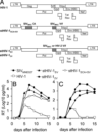
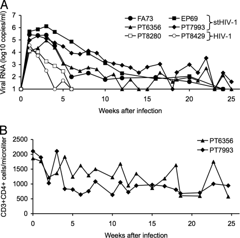
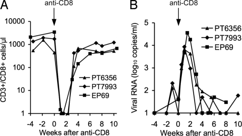
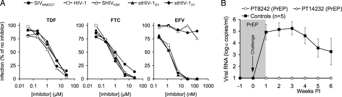
The lack of a primate model that utilizes HIV-1 as the challenge virus is an impediment to AIDS research; existing models generally employ simian viruses that are divergent from HIV-1, reducing their usefulness in preclinical investigations. Based on an understanding of species-specific variation in primate TRIM5 and APOBEC3 antiretroviral genes, we constructed simian-tropic (st)HIV-1 strains that differ from HIV-1 only in the vif gene. We demonstrate that such minimally modified stHIV-1 strains are capable of high levels of replication in vitro in pig-tailed macaque (Macaca nemestrina) lymphocytes. Importantly, infection of pig-tailed macaques with stHIV-1 results in acute viremia, approaching the levels observed in HIV-1-infected humans, and an ensuing persistent infection for several months. stHIV-1 replication was controlled thereafter, at least in part, by CD8+ T cells. We demonstrate the potential utility of this HIV-1-based animal model in a chemoprophylaxis experiment, by showing that a commonly used HIV-1 therapeutic regimen can provide apparently sterilizing protection from infection following a rigorous high-dose stHIV-1 challenge.


Conflict of interest statement

The authors declare no conflict of interest.

Figures

Fig. 1.
Fig. 1.
Replication of stHIV-1 variants in pig-tailed macaque lymphocytes in vitro. (A) Schematic representation of the viral constructs used in this study, shaded regions indicate sequences from HIV-2ROD or SIVMAC239, as indicated. All HIV-1-derived constructs used the macaque-adapted HIV-1 envelope from SHIVKB9. (B, C) RT activity in culture supernatants (expressed as log10 pg/ml) was measured every 2 or 3 days for 15 days following inoculation of pig-tailed macaque lymphocytes with the indicated viruses. The 2 graphs shown in (B) and (C) represent independent experiments employing 2 different pig-tailed macaque donors.
Fig. 2.
Fig. 2.
Infection of pig-tailed macaques with HIV-1 and stHIV-1. (A) Four macaques were infected with a 1:1 mixture of stHIV-1SV and stHIV-12V (filled symbols) and 2 macaques were infected with the same dose (2 × 10⁶ i.u.) of HIV-1 (open symbols). Plasma viremia was measured by RT-PCR and is given as the log10 viral RNA copies per milliliter of plasma. (B) CD4+ T cells, enumerated using FACS analyses in 2 of the 4 stHIV-1-infected macaques over the course of infection. CD4 counts in the 2 other macaques were monitored on a different schedule (see SI Text, Fig. S1).
Fig. 3.
Fig. 3.
Antibody responses to stHIV-1 infection in pig-tailed macaques. Western blot analysis using plasma recovered from the 4 stHIV-1-infected macaques at the indicated time points following stHIV-1 infection. Serum from an HIV-1-infected human long-term nonprogressor was used as a positive control.
Fig. 4.
Fig. 4.
Effect of depletion of CD8+ cells on stHIV-1 replication in vivo. (A) Effect of anti-CD8-antibody infusion on the numbers of CD8+ T cells in peripheral blood, as determined by FACS analyses. Note that analysis of lymph node biopsies at 1 week after antibody infusion revealed that 0%, 8%, and 0.1% of the CD3+ cells were CD8+ for animals PT6356, PT7993, and EP69, respectively. (B) Plasma viremia, measured by RT-PCR in the weeks preceding, during, and following CD8+ T-cell depletion in 3 animals.
Fig. 5.
Fig. 5.
Antiretroviral drug prophylaxis of stHIV-1 infection. (A) Sensitivity of SIVMNE027, SHIVKB9, HIV-1, and stHIV-1 strains to inhibition by the nucleoside (TDF and FTC) and non-nucleoside (EFV) RT inhibitors, that comprise a mixture commonly used for treatment of HIV-1 infection. TZM cells were infected with each virus in the presence of the indicated concentrations of each drug, and the level of infection is plotted as the percentage of that in the absence of drug. (B) Two macaques received chemoprophylaxis with a TDF/FTC/EFV combination (PrEP) and were challenged intravenously, at 1 week after prophylaxis initiation, with the same dose of stHIV-1SV/2V as was used in Fig. 2. Treatment was continued for 1 week following the challenge. A contemporaneous control macaque was challenged but untreated. Plasma viremia was measured for the ensuing 5 weeks, using RT-PCR as in Fig. 2, and compared to the 4 previously inoculated and the contemporaneous control for which the mean and standard deviation of the plasma viremia over the first 6 weeks of infection is plotted.

References

    1. Ambrose Z, KewalRamani VN, Bieniasz PD, Hatziioannou T. HIV/AIDS: in search of an animal model. Trends Biotechnol. 2007;25:333–337. - PubMed
    1. Fultz PN. Nonhuman primate models for AIDS. Clin Infect Dis. 1993;17(Suppl 1):S230–S235. - PubMed
    1. Daniel MD, et al. Isolation of T-cell tropic HTLV-III-like retrovirus from macaques. Science. 1985;228:1201–1204. - PubMed
    1. Reimann KA, et al. A chimeric simian/human immunodeficiency virus expressing a primary patient human immunodeficiency virus type 1 isolate env causes an AIDS-like disease after in vivo passage in rhesus monkeys. J Virol. 1996;70:6922–6928. - PMC - PubMed
    1. Harouse JM, Gettie A, Tan RC, Blanchard J, Cheng-Mayer C. Distinct pathogenic sequela in rhesus macaques infected with CCR5 or CXCR4 utilizing SHIVs. Science. 1999;284:816–819. - PubMed

Publication types

Substances