. 2009 Mar 17;106(11):4513-8.
doi: 10.1073/pnas.0807974105. Epub 2009 Mar 2.

Hydrogen sulfide as a mediator of human corpus cavernosum smooth-muscle relaxation

Affiliations

Hydrogen sulfide as a mediator of human corpus cavernosum smooth-muscle relaxation

Roberta d'Emmanuele di Villa Bianca et al. Proc Natl Acad Sci U S A. .

Abstract

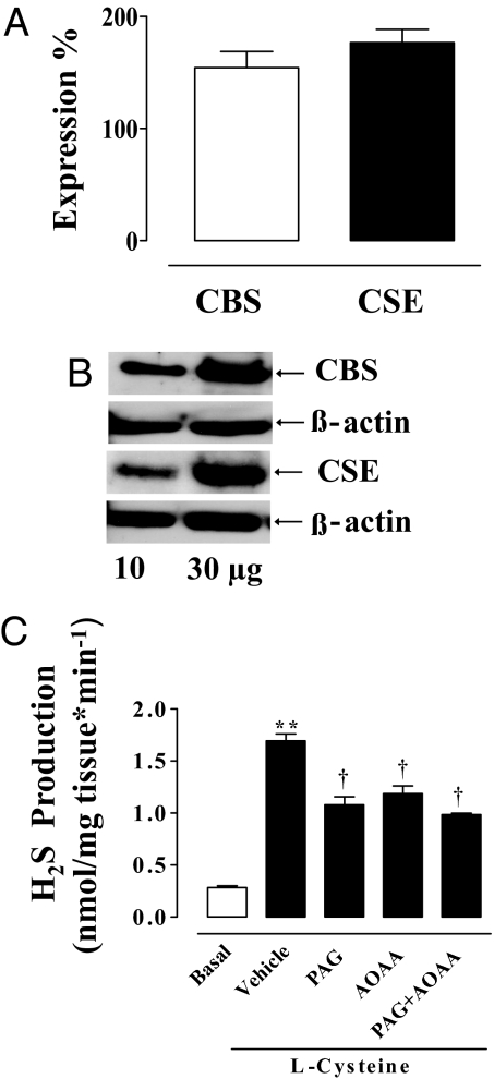
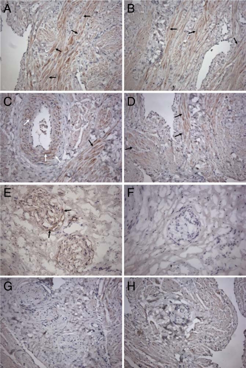
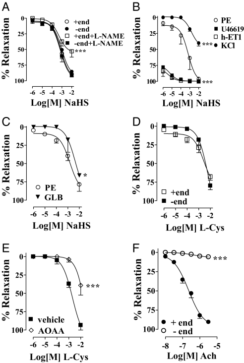
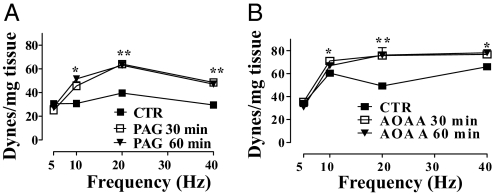
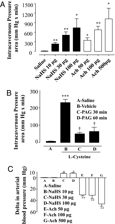
Hydrogen sulfide (H(2)S) is synthesized by 2 enzymes, cystathionine beta-synthase (CBS) and cystathionine gamma-lyase (CSE). L-Cysteine (L-Cys) acts as a natural substrate for the synthesis of H(2)S. Human penile tissue possesses both CBS and CSE, and tissue homogenates efficiently convert L-Cys to H(2)S. CBS and CSE are localized in the muscular trabeculae and the smooth-muscle component of the penile artery, whereas CSE but not CBS is also expressed in peripheral nerves. Exogenous H(2)S [sodium hydrogen sulfide (NaHS)] or L-Cys causes a concentration-dependent relaxation of strips of human corpus cavernosum. L-Cys relaxation is inhibited by the CBS inhibitor, aminoxyacetic acid (AOAA). Electrical field stimulation of human penile tissue, under resting conditions, causes an increase in tension that is significantly potentiated by either propargylglycine (PAG; CSE inhibitor) or AOAA. In rats, NaHS and L-Cys promote penile erection, and the response to L-Cys is blocked by PAG. Our data demonstrate that the L-Cys/H(2)S pathway mediates human corpus cavernosum smooth-muscle relaxation.


Conflict of interest statement

The authors declare no conflict of interest.

Figures

Fig. 1.
Fig. 1.
CBS and CSE: activity, Western blot analysis, and qRT-PCR of human penile tissue. (A) HCC-expressed mRNA for both CBS and CSE as determined by qRT-PCR. (B) Representative Western blot analysis for CBS and CSE. (C) HCC homogenate produced H2S under basal conditions (open bar). Incubation of HCC homogenate with 10 mM l-Cys caused a significant increase in the H2S production compared with basal values (**, P < 0.001). PAG (10 mM), 1 mM AOAA, or 10 mM PAG plus 1 mM AOAA significantly inhibited the l-Cys-induced increase in H2S production (†, P < 0.01). Data represent the mean ± SEM from 3 or 4 different human specimens.
Fig. 2.
Fig. 2.
Immunochemistry for CBS and CSE in HCC. (A–D) Immunohistochemical detection of CSE and CBS in HCC tissue. Immunoreactivity and nuclear staining appear brown (DAB) and blue (hematoxylin counterstain), respectively. CSE was detected in trabecular muscular tissue (A and B, black arrows) and vascular smooth-muscle cells (C, white arrows). Immunoreactivity for CBS was mostly observed in trabecular muscular tissue (D, black arrows). Results illustrated are from a single experiment and are representative of 3 different specimens. (Original magnification, 200×.) (E–H) Immunohistochemical detection of CSE and CBS in HCC nerve fibers. Immunoreactivity and nuclear staining appear brown (DAB) and blue (hematoxylin counterstain), respectively. CSE was detected in nerve fibers in cryostat (E, arrows) and not in paraffin (G) sections. Both cryostat (F) and paraffin (H) sections lacked immunoreactivity for CBS. Results illustrated are from a single experiment and are representative of 3 different specimens. (Original magnification, 200×.)
Fig. 3.
Fig. 3.
NaHS or l-Cys effect on HCC strips. (A) NaHS-relaxed HCC strips with (+end) or without (−end) endothelium; 100 μM l-NAME inhibited NaHS-induced relaxation at higher concentrations tested and in the presence of endothelium only (***, P < 0.0001). (B) HCC strips without endothelium precontracted with 10 nM U46619 or 30 nM h-ET1 displayed a significantly increased relaxant response to NaHS compared with 3 μM PE (***, P < 0.0001). NaHS-induced relaxation in HCC strips without endothelium contracted with 80 mM KCl was significantly reduced compared with 3 μM PE contractions (***, P < 0.0001). (C) Incubation of HCC with 150 μM glibenclamide (GLB) before 3 μM PE reduced NaHS-induced relaxation (*, P < 0.05). (D) l-Cys relaxed HCC strips precontracted with 3 μM PE with (+end) or without (−end) endothelium. (E) One millimolar AOAA inhibited l-Cys-induced relaxation in HCC strips without endothelium (−end) precontracted with 3 μM PE (***, P < 0.0001). (F) Ach caused relaxation of HCC strips with (+end) but not without (−end) endothelium precontracted with 3 μM PE (***, P < 0.0001). Experiments were performed on 8 strips for each of the NaHS experiments, 5 strips for each of the l-Cys experiments, and 15 strips for the Ach experiment.
Fig. 4.
Fig. 4.
EFS of HCC strips. EFS caused a frequency-related increase in basal tone. Incubation of HCC strips with 10 mM PAG for 30 or 60 min (A) or with 1 mM AOAA for 30 or 60 min (B) significantly increased the EFS-induced contraction [*, P < 0.05; **, P < 0.01 vs. control (CTR)]. Tissue responses to EFS are expressed as force in dynes per milligram of tissue. Experiments were performed on 3 different specimens.
Fig. 5.
Fig. 5.
Effect of NaHS or l-Cys on intracavernous pressure in anesthetized rats. (A) NaHS (10, 30, and 100 μg per rat) caused significant dose-dependent increases in intracavernous pressure. Ach (50, 100, and 500 μg/ per at) caused a dose-dependent increase in intracavernous pressure. (B) l-Cys at a dose of 30 μg per rat caused an increase in intracavernous pressure. Intravenous administration of 50 mg/kg PAG significantly inhibited l-Cys-induced penile erection (***, P < 0.0001; †, P < 0.05). (C) NaHS at a dose of 10 μg per rat did not cause any change in arterial blood pressure whereas 30 and 100 μg of NaHS per rat caused a significant increase in blood pressure. Ach (50, 100, and 500 μg per rat) caused a dose-dependent significant decrease in blood pressure (**, P < 0.01; *, P < 0.05 vs. saline). Data were obtained from 7 separate experiments for NaHS and 4 for l-Cys.

References

    1. Li L, et al. Hydrogen sulfide is a novel mediator of lipopolysaccharide-induced inflammation in the mouse. FASEB J. 2005;19:1196–1198. - PubMed
    1. Zhong G, Chen F, Cheng Y, Tang C, Du J. The role of hydrogen sulfide generation in the pathogenesis of hypertension in rats induced by inhibition of nitric oxide synthase. J Hypertens. 2003;21:1879–1885. - PubMed
    1. Bhatia M, et al. Role of hydrogen sulfide in acute pancreatitis and associated lung injury. FASEB J. 2005;19:623–625. - PubMed
    1. Zhao W, Zhang J, Lu Y, Wang R. The vasorelaxant effect of H2S as a novel endogenous gaseous KATP channel opener. EMBO J. 2001;20:6008–6016. - PMC - PubMed
    1. Wang R. Two's company, three's a crowd: Can H2S be the third endogenous gaseous transmitter? FASEB J. 2002;16:1792–1798. - PubMed

LinkOut - more resources